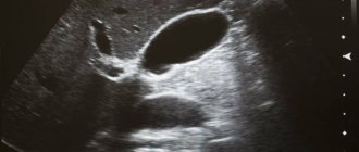
MRI of the lungs is one of the most informative and safe procedures that allows you to get a clear picture of the current condition of the patient’s body. The technology helps to identify pathologies at an early stage and track noticeable changes in tissue structure.
Due to the absence of harmful effects on the body, tomography can be performed several times. This helps to track the dynamics of treatment and timely adjust the list of medications or therapy.
There are minimal contraindications to MRI of the lungs. The method can even be used for pregnant women and children. At the same time, it is possible to obtain the clearest and most informative images and high-quality three-dimensional images.
In this material we will consider indications for the examination, methods of preparation for MRI of the lungs in adults, contraindications and other important aspects of visiting the center. This way you will get a complete idea of what you should pay attention to when making an appointment with our specialists.
General characteristics of the examination
Do they do MRI of the lungs and bronchi? For accurate diagnosis of diseases of the lungs and bronchi, in addition to ultrasound, CT, fluorographic examination
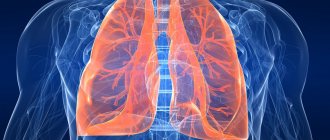
, patients undergo MRI. It shows whether there are tumors, swelling in these organs, whether blood circulation is impaired in them, and also allows you to quickly and effectively provide assistance. What is an MRI of the lungs? It is a photo (layer-by-layer) of the structures of the organ being examined, giving a complete picture of the disease. MRI also helps to assess the patient’s respiratory function, determine the presence of thickenings in the walls of the lungs and bronchi, and understand the causes of the disease.
CT scan for oncology
CT - computed tomography is another way to diagnose cancer tumors. The method is based on the ability to obtain images using X-rays.
The advantages of CT are as follows:
- High accuracy of the results obtained and three-dimensional image.
- Tomography takes a couple of minutes.
- There are no contraindications that MRI has.
- The dose of X-rays is lower than in an X-ray machine.
- The cost of a CT scan is lower than the cost of an MRI.
CT scanning is somewhat similar to MRI. The scanner is different in that it has the shape of a donut, it is narrower than an MRI scanner, there are no closed spaces, and people with claustrophobia will feel comfortable.
Each of these procedures is absolutely painless and safe; there are no age restrictions for its implementation, which means that any such tomography can be performed even on infants. It is hardly possible to distinguish the differences between the procedures, since these are completely different devices; the differences lie only in contraindications, indications and methods of influencing the human body.
Indications for the procedure
Indications for MRI of the lungs and bronchi are:
- various types of bronchitis, including dust, infectious, eosinophilic, diffuse, terry and others;
- bronchiolitis obliterans;
- bronchial tuberculosis;
- pneumonia;
- bronchopneumonia;
- cystic fibrosis (childhood disease);
- bronchial hyperactivity;
- bronchial edema;
- carcinoidbronchus.
Similar diseases have similar symptoms. These are shortness of breath, attacks of dry cough, hemoptysis, pain in the chest, increased body temperature and others. To accurately determine the diagnosis, as practice shows, patients are often prescribed MRI of the lungs and bronchi, which helps in the future to identify the exact cause of the disease and prescribe effective therapy.
Dust bronchitis
Dust bronchitis is a non-infectious lesion of the bronchi, but a consequence of the mechanical or chemical effect on them of various dust particles in the surrounding air. This disease is occupational in nature and almost always becomes chronic. Dust bronchitis is often diagnosed in miners, miners, construction workers, workers in flour mills, textiles or woodworking enterprises.
Bronchial tuberculosis
Bronchial tuberculosis most often becomes a consequence of actively developing tuberculosis of the lungs, larynx, and trachea. It is an ulcerative, fistulous or infiltrative lesion of the bronchial cavity. Bronchial tuberculosis can occur due to:
- germination of granulations from previously infected lymph nodes in the walls of the bronchi;
- discharge of infected sputum through the walls of the bronchi;
- dispersion of bacteria along the lymphatic tract;
- spread of bacteria through the blood vessels of the bronchi.
Bronchiolitis obliterans
Bronchiolitis obliterans is a lesion of the small human airways – bronchioles. The occurrence of this disease can be provoked by: cytomegalovirus, HIV infection, mycoplasma infection, lupus erythematosus, and intestinal inflammation. Also, bronchiolitis can be a consequence or complication after a complex heart and lung transplant, bone marrow transplant, after taking certain medications, or undergoing radiation therapy.
Infectious bronchitis
Infectious is the most common type of bronchitis. It is characterized by damage to the lower respiratory tract. The causative agents can be fungus, virus, bacteria. Infectious bronchitis often occurs as a complication of influenza or ARVI.
Significant symptoms of infectious bronchitis are:
- dry wheezing when listening to the patient's breathing;
- hard breath;
- dry cough;
- chest pain;
- elevated temperature up to 38-39℃;
- general weakness.
Treatment regimens for bronchitis are very diverse, so it is very important to determine the cause of the disease, its severity, as well as the presence of concomitant diseases. MRI in such cases should provide comprehensive information.
Features of the use of contrast during MRI of the lungs
In order to increase the level of clarity of the resulting image, doctors often use additional contrast enhancement. A special drug containing a high content of gadolinium salts is injected intravenously.
It is the use of contrast enhancement that makes it possible to find tumors in the early stages and whether there are signs of a pathological process in the body where they are concentrated.
The drug used does not pose a danger to the human body. A minimum of time is spent on its removal - usually within a day all components are removed from the body naturally.
When using a contrast agent, the duration of the procedure increases by one and a half to two times. But this helps to achieve maximum information, accurately diagnose and begin treatment.
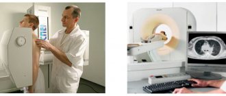
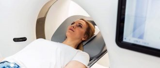
How is the procedure done?
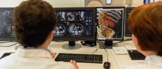
How an MRI of the lungs is done: the video is sent to the doctor’s computer. It consists of thin sections of the organ being examined. The average duration of an MRI procedure of the lungs and bronchi is 20 minutes. During this time, the movable table gradually moves along the tomograph, which scans the patient’s body and takes pictures of the organs being examined in several projections and from different angles. During the procedure, the patient lies motionless. He can only hear noise, no unpleasant sensations or discomfort are felt. Immediately after the procedure, ready-made images are obtained, and a specialist can decipher them. The correct diagnosis based on tomography results is made by the attending physician.
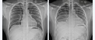
What does MRI of the lungs and bronchi show?
When a doctor conducts an examination on a tomograph, he receives a three-dimensional image and high-quality images, which can also be recorded on electronic media. Upon careful study of the obtained indicators, a detailed conclusion is drawn up based on the results.
There are several main indicators that can be studied based on the results obtained:
- The size of the lungs, the clarity of the contours.
- The degree of patency of blood vessels.
- The presence or absence of fluid inside the lungs.
- Signs of development of various neoplasms.
- Blackouts.
- Dilatation and patency of the bronchi.
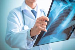
All pathologies will be visible in the image. They appear as darkening or very light areas that need further examination.
Preparation for MRI of the throat (contraindications)
MRI of the throat does not require any preliminary manipulations. Before performing an MRI of the larynx, the doctor must ensure that there are no contraindications. They are:
- The patient's age is less than 7 years. It is believed that children do not have proper perseverance and will not be able to behave calmly during the procedure.
- Early pregnancy (first trimester). At this time, there is a small chance of harm to the fetus. To completely eliminate the risk, other methods are used.
- Allergies and kidney failure. They become contraindications when a contrast agent is used during MRI.
- Presence of pacemakers or metal prostheses.
Before the study, you need to get rid of metal objects and things that could be damaged by the magnetic field. They are:
- Watches, piercings, coins.
- Bank plastic cards.
- Electronic devices, such as a mobile phone.
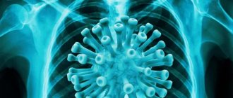
Symptoms of lung cancer
Signs of an oncological process are nonspecific, as they are observed in many pathologies. Symptoms appear when the tumor spreads into the bronchial mucosa. The first symptom of lung cancer may be a dry, persistent cough. The central form has a clear clinical picture:
- cough clears up quickly;
- hemoptysis occurs;
- shortness of breath with minimal activity;
- low-grade fever;
- general malaise, weakness.
The face becomes puffy, acquiring a cyanotic (blue) tint to the skin. This is due to impaired lymphatic drainage.
When metastases of lung cancer appear, damage to internal organs is observed. Malignant cells are spread throughout the body through the bloodstream, so the brain, adrenal glands, and liver can be involved.
See what is hidden: from ultrasound to CT
Their names are always well-known: a neighbor was prescribed an MRI yesterday, a friend urgently needs an X-ray, and only kindergarten students have not heard of fluorography.
However, knowledge, as a rule, does not extend beyond terminology.
For the average citizen, it remains unclear when one method or another is used, and what its advantages and disadvantages are.
Let's fill in the gaps.
Ultrasound - simply from the first days of life
Ultrasound, or ultrasonography, is an excellent diagnostic tool for studying the anatomical features and functioning of many organs and systems in real time.
How to sign up for diagnostics
You can book the type of scan you need or call a doctor at home to conduct a coronavirus test through our free service.

On the main page, indicate the name of the procedure, the area where the clinic is located, and examine the list of organizations that opens. If necessary, you can use other search filters. To make an appointment, select a medical center, call the consultation service staff and book a session at a convenient time with discounts.
Sources used:
- Gedymin L.E., Filippov V.P., Evgushchenko G.V., et al. The role of biopsy in the diagnosis of pulmonary diseases at the prehospital level of observation // Clinical Medicine. - 2009. - No. 4. — P.41-44.
- Ivanova E.V., Bilichenko T.N., Chuchalin A.G. Morbidity and mortality of the working age population in Russia due to respiratory diseases in 2010 -2012. // Pulmonology. - 2015. - T. 25. - No. 3. - P.291-297.
- Karashchuk N.P., Kiseleva M.V. Lung cancer and tuberculosis // Scientific Medical Bulletin of Ugra. -2014. — No. 1-2(5-6). — P.71-73.
- Kartashov M.V., Kartashova O.M., Kotlyarov P.M. First experience of using diffusion-weighted magnetic resonance imaging for small cell lung cancer // Medical visualization. – 2011. – No. 4. – P.28-33.
- Kotlyarov P.M. Post-processing of multislice computed tomography data in the refined diagnosis of pathological changes in diffuse lung diseases // Pulmonology. - 2021. - No. 4. — P.472-477.
- Kotlyarov P.M., Sergeev N.I. Radiation research methods in the differential diagnosis of parasitic and tumor lesions of the lungs // Siberian Journal of Oncology. – 2021. – No. 15(4). – P.33-39.
- Ternovoy S.K., Nasnikova I.Yu., Morozov S.P. Modern computed tomography in clinical medicine // Kremlin Medicine. Clinical Bulletin. - 2008. - No. 2. — P.9-13.
- Tyurin I.E. Differential diagnosis of single lesions in the lungs // Polyclinic. - 2014. - No. 3-1. — P.28-32.
- Yudin A.L., Afanasyeva N.I., Abovich Yu.A., et al. High-resolution computed tomography in the diagnosis of interstitial pneumonia // Medical visualization. -2002. - No. 4. — P.40-48.
How is magnetic resonance imaging of the OGK performed with contrast?
Diagnosis of the chest organs using innovative high-power MRI scanners is gaining popularity in Europe. Some Western scientists propose replacing mass fluorography with MRI. Examination of the lungs in this way eliminates tissue irradiation, which is the most negative factor of many radiation methods.
Intravenous administration of a contrast agent enhances the visibility of blood vessels. After saturating the arteries with an enhancing substance containing a paramagnetic compound, visibility improves significantly, which makes it possible to identify a number of nosologies:
- Constrictions;
- Violation of anatomical location;
- Blockade by atherosclerotic plaque;
- Embolic areas;
- Deposition of blood clots;
- Congenital and acquired anomalies.
Modern equipment is equipped with special syringe dispenser systems for conducting basal-bolus contrast. The procedure involves the parallel administration of gadolinium-based substances and the study of tissues on tomograms. The image quality is determined by the scanning step. The choice of a short scanning step (several millimeters) improves visibility. High-field equipment has three-dimensional reconstruction modes to create a spatial model of the object under study.
Before the procedure, it is necessary to exclude allergic reactions to the main active ingredient of the contrast compound - gadolinium. Doctors analyze the patient’s outpatient card and conduct a provocative test. Only in the absence of a health condition can the procedure be performed.