The pituitary gland is located in the central sections of the base of the skull. Despite its small size (~ 6x15 mm) and weight (less than 1 gram), the pituitary gland is an important gland of the body. The pituitary gland produces hormones and ensures the functioning of the endocrine system. The anterior lobe of the pituitary gland produces hormones responsible for the development, growth, and stimulation of the endocrine glands. The posterior lobe produces hormones that stimulate smooth muscle and affect the (excretory) function of the kidneys.
The pituitary gland produces growth hormone; it also regulates the activity of the central nervous system, so a person’s mood depends on its work. The pituitary gland is responsible for maternal instincts and influences the process of lactation (milk production in the female body). Even the smallest changes can cause serious disruptions in the body's functioning. MRI of the pituitary gland is used to detect these disorders in the early stages.
In what cases is an MRI of the spine needed?
There are the following indications for MRI of the spine:
- intercostal neuralgia;
- dizziness, headaches;
- discomfort in the neck or lower back;
- pain in the upper or lower extremities, limitation of their mobility;
- previous spinal injuries;
- tinnitus, hearing loss;
- blurred vision and other manifestations of cerebral circulation pathologies;
- numbness or complete loss of mobility of the limbs;
- loss of consciousness;
- painful sensations in the back, the nature of which is not established.
MRI of the spine is prescribed for other clinical symptoms that indicate pathological processes in the spinal structures, as well as if previously performed studies cannot unambiguously determine the nature of the disease.
Thanks to MRI, the following pathologies can be diagnosed:
- compression or inflammation of the nerves;
- infectious processes;
- consequences of previous injuries, in particular, fractures, bruises, compression, etc.;
- degenerative-dystrophic processes of intervertebral discs;
- spinal cord compression;
- multiple sclerosis;
- disorders of the spinal cord and its membranes (myelitis, arachnoiditis);
- spondylosis, spondyloarthrosis, osteochondrosis;
- narrowing of the spinal canal;
- osteoporosis;
- neoplasms of various nature;
- compression of the vertebral arteries and other vascular disorders.
MRI also detects acquired and congenital defects in the development of spinal structures.
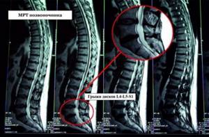
It should be noted that MRI is the “gold” standard for diagnosing intervertebral hernias at the earliest stages of their development, which ensures the most effective therapy.
MRI with or without contrast
MRI of the spine with contrast is required for the differential diagnosis of space-occupying lesions, as well as to identify certain vascular pathologies, such as malformations. Due to the fact that tumor processes have a dense network of blood vessels, they are well contrasted, unlike the intervertebral disc, which does not accumulate contrast. When detecting osteomyelitis and metastatic lesions of the vertebrae, fat suppression MR mode is used.
Contrast MRI of the spine allows you to determine the exact size and stage of tumor development, differentiate benign and malignant formations, determine the characteristics of the relationship with neighboring tissues - germination, etc.
The need for contrast, as well as the amount of the drug, is determined by the attending physician or the doctor in the MRI room.
What does an MRI of the pituitary gland show?
Magnetic resonance imaging provides a clear image of the structures of the pituitary gland, which shows the venous and arterial vessels. Medical equipment uses high-quality equipment that allows you to obtain high-quality images. MRI of the pituitary gland in St. Petersburg is a harmless and painless procedure with which you can determine:
- cysts;
- tumors;
- developmental anomalies;
- ease of execution - the main part of the study is automated;
- does not require complex patient preparation;
- minimum number of contraindications.
Preparation for the procedure
As a rule, special preparation for MRI of the pituitary gland is not required. View all information about preparing for the procedure. The price for MRI of the pituitary gland in St. Petersburg is indicated in our price list.
Before carrying out the procedure, study the contraindications.
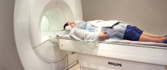
How is an MRI of the spine performed?
The study is no different from other types of MRI scans. The patient is placed on the moving table of the tomograph, and a radiofrequency coil is placed on the area of the spine being examined, which helps obtain accurate images. The table is then moved into the machine so that the area of interest is located in the middle of the scanner, and scanning begins. During operation, the device makes loud sounds, so the patient is given earplugs or headphones. If the patient experiences discomfort during the examination, he can contact the medical staff using the communication system equipped with the tomograph.
How to prepare for an MRI of the spine
Patients often ask, is it possible to eat before an MRI of the spine? If you have a non-contrast MRI, no preparation is required - you can take food, drinks, and medications.
Preparing for a tomography with contrast involves refusing to eat 2 hours before the scan, since one of the side effects of the contrast drug is nausea and vomiting.
What to take for research? The patient needs to take a passport, a doctor’s referral, if available, and, if available, the results of previous diagnostics - CT scan, x-ray, etc.
What to wear? Due to exposure to a magnetic field during scanning, clothing should not have metal elements. Before the examination itself, the patient is required to remove the phone, watch and other ferromagnetic accessories. Please note that most medical centers provide disposable medical clothing for the duration of the study.
How long does a spine MRI procedure take?
The answer to the question of how long it takes to do an MRI of the spine depends on the area of study, the need for contrast and the provision of anesthesia.
An MRI of one part of the spine usually lasts about 15-20 minutes. If you need to scan two or three areas, then this time increases proportionally.
MRI with contrast usually takes 15 minutes longer than a native scan.
If the patient suffers from claustrophobia, uncontrollable convulsive syndrome, with severe pain that does not allow the patient to maintain a stationary position for a long time, he needs to be provided with medicated sleep. In this case, the MRI takes 15-20 minutes longer, and after the scan is completed, the patient must remain in the medical facility for some time to monitor his condition.
Description of MRI of the spine
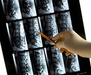
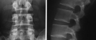
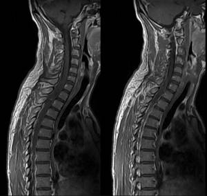
After the scan, the doctor receives a series of thin, layer-by-layer black-and-white MRI images of the spine. Based on a comparison with the norms of density, homogeneity, shape and size of spinal structures, and the presence of foreign bodies, a conclusion is made about the presence of any pathology. The specialist evaluates the narrowing of the bony spinal canal, the presence of marginal bone growths on the surfaces of the vertebral bodies, the structure and location of the vertebrae, thickening of the posterior longitudinal and other ligaments, the state of lordosis and other parameters.
Please note that MRI scans of the spine should be read only by radiologists, and not by the patient himself, in order to avoid misinterpretation.
Consequences of MRI of the spine
Unlike X-ray examination methods, there is no harm from MRI due to the lack of radiation exposure to the body. The use of a strong uniform magnetic field in combination with radio frequency radiation is absolutely safe for diagnosing the spine in patients of any age, including newborns.
Is it harmful to have an MRI of the spine with contrast? The contrast used in MRI does not contain iodine compounds and is non-toxic. Rarely, it can cause adverse reactions of a local and general nature, manifested in tachycardia, drowsiness, headaches, nausea, etc. In the vast majority of cases, it is well tolerated and does not cause any discomfort to the patient.
The question of the harmfulness of MRI also depends on contraindications to the procedure. If the patient has a pacemaker, cerebral vessel clips, or ferromagnetic implants, an MRI is strictly forbidden. If contrast is necessary, the excretory function of the kidneys, the condition of the liver and the cardiovascular system are assessed, since if they are severely impaired, the possibility of contrast MRI is determined by doctors.
Is it worth doing an MRI of the spine?
MRI has the highest degree of reliability in studying the condition of the spine in comparison with other techniques. If there is a suspicion of post-traumatic changes, intervertebral hernias, vascular disorders, degenerative or tumor processes, it is definitely worth performing an MRI of the spine. However, it is better to consult with a specialized doctor to determine the exact scanning mode and area before MRI.
Indications and contraindications for MRI of the lumbosacral spine
Indications for MRI of the lumbosacral area are recovery from spinal injuries, “weakness” of the bladder, and patient complaints of back and lower back pain. The procedure may also be prescribed if tumor diseases are suspected.
Although MRI is generally the safest type of medical diagnostics at the moment, it still has its own characteristics and contraindications:
- For patients suffering from claustrophobia, it is important to use an open tomograph model. If the most common option in the form of a pipe is used, it is recommended to take sedatives.
- As a rule, the tomograph is designed to weigh up to 120 kg. There are models for patients weighing up to 180 kg.
- Breastfeeding should not be performed after an MRI with contrast agent for 48 hours. During pregnancy, MRI of the lumbosacral region is not prescribed, since there is currently no accurate data on the effects of electromagnetic waves on the fetus, and contrast agents can penetrate the placenta.
- Individual intolerance to the contrast agent and allergic reactions are possible. In addition, in cases of kidney failure, contrast tends to accumulate in the body. Both cases are contraindications for contrast MRI.
- If you have a pacemaker, ferromagnetic implants or fragments, MRI is absolutely contraindicated. If a patient has a colored tattoo in the lumbar region, burns are possible when exposed to a magnetic field, since the dyes contain metal compounds.
- ·The procedure lasts about half an hour, so it is not prescribed for acute injuries, for urgent diagnosis, and in cases where pain in the spine does not allow the patient to lie on his back motionless the entire time. Children under the age of seven are usually not prescribed an MRI, since it is psychologically difficult for a small child to spend such a long time without moving. In these cases, computed tomography (CT) is used to replace this type of diagnosis.
Get an MRI of the pituitary gland in St. Petersburg
Magnetic resonance imaging of the pituitary gland can be done at Medical in St. Petersburg. The procedure helps in identifying pathologies. The diagnostic cost includes:
- Examination on a modern tomograph Vantage Elan 1.5T (manufactured by Canon, Japan), which belongs to the class of high-field tomographs of the latest generation, providing safe, reliable and high-quality research.
- A detailed conclusion made based on the images by a radiologist. The description of the study is checked by the chief physician.
- Internal research quality control
- 100% guarantee of photo quality
- Detailed information about prices can be found in the “Prices” section.
- You can view current promotions in the “Promotions” section.
The procedure lasts 30 minutes, you can sign up for a convenient time on any day of the week. The medical department uses modern equipment, which ensures high quality images.
How to prepare for a lumbar MRI
The procedure does not require special preparation. From the package of documents you will need the results of previous examinations, if any, and a doctor’s referral, since this allows you to focus the work of the diagnostician on the truly problem area.
Before the procedure itself, you should visit the toilet and get rid of all metal objects both on clothing and on the body - they create interference and spoil the quality of the image.
If the patient has a fear of closed spaces or any mental disorders, you should inform the doctor about this in advance. Then he will prescribe a mild sedative.
Contraindications for the procedure
- The presence of electronic devices and metal objects in the body (stents, knitting needles and staples in joints, pacemaker, insulin pump, fixed dentures). When exposed to a magnetic field, there is a risk that devices will break. If the metals of foreign objects are non-magnetic, then the procedure is allowed.
- Claustrophobia (during the procedure, the patient is placed inside a tomograph that looks like a closed tunnel).
- Large body weight (the equipment has a weight and circumference limit of 120 kg and 150 cm).
Normal MRI indicators of all parts of the spine
MRI of the spine: normal and pathological
The diagnostician draws attention to the average population statistical parameters that are accepted as the norm: the absence of pathological curvatures of the spinal column, spinal cord compression, focal formations, edema, hernial protrusions, metastatic lesions, preservation of bone structures, etc. All changes will be reflected in detail in the protocol research. The final diagnosis is made by the attending physician. In doubtful cases, MRI results are assessed collectively. You can get a “second opinion” if you send the images for consultation to a specialized medical center. This service is currently provided by many large Russian and foreign clinics.
Types of MRI depending on the device
MRI examination of the spine is carried out using open and closed type devices.
Closed-type devices are high-field; examination with them is more accurate due to the greater strength of the magnetic field (from 1.5 Tesla). The cost of scanning increases with the power of the tomograph, since the highest field scanners are the most expensive to maintain. Such devices, even without contrast enhancement, are capable of visualizing formations up to 2 mm, which is extremely important when searching for metastases, foci of multiple sclerosis, etc. However, in addition to the high cost, examination with such tomographs is often not comfortable enough for patients. The narrow tunnel of the device limits examination for obese patients and for people suffering from claustrophobia.
Examination with a low-field tomograph is more comfortable due to the free lateral space. Despite the low induction force (up to 0.5 Tesla), the power of such scanners is quite sufficient for a full study of most pathologies (exceptions are oncological processes, diseases of the spinal cord, and conditions after spinal surgery).
A variety of open scanners are polyposition tomographs, which allow MRI of the spine to be performed while standing, sitting, or tilted. It is believed that the vertical position is more desirable for examining the spine and some organs (kidneys, heart), since this position shows the true position of the tissues under the influence of gravity.
Dangerous "finds"
MRI of the peritoneum and retroperitoneal space allows us to identify diseases that occur in a latent form (that is, they do not manifest symptoms and do not bother the patient).
- Stones in the bile ducts. Gallstones are a dangerous disease that never goes away on its own and requires long-term treatment or surgery. But as long as they lie “quietly” in the gallbladder, that’s not so bad. It is much worse when the stone begins to migrate and gets stuck in the bile ducts. MR imaging of the peritoneum can often identify such an unexpected finding.
- Adhesive disease. The disease appeared along with surgery and will, apparently, exist as long as the surgical treatment itself. The pathology is postoperative adhesions in the peritoneum and is found in 65-95% of patients who have undergone abdominal surgery (not only abdominal, but also minimally invasive endoscopic). There are also known cases of adhesive disease caused by inflammation of the abdominal organs. Against the background of fibrous cords (adhesions), endometriosis, intestinal obstruction, and deformation of neighboring organs can develop. Therefore, it is important to detect pathology and prevent its dangerous consequences.
MRI of the abdominal cavity is one of the most informative methods for diagnosing various diseases that can be asymptomatic, painful, or life-threatening.