Cholesterol is a fatty organic substance that is part of cell membranes and is necessary for the functioning of every cell in the body. However, excess cholesterol in the body can be dangerous, as it is considered one of the causes of the development of atherosclerosis and cardiovascular diseases: coronary heart disease, heart attack, stroke, etc.
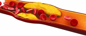
Most of the cholesterol necessary for the functioning of the body is synthesized in the liver, but it should be remembered that it also enters the body with food. In combination with genetic predispositions and physiological characteristics of the body, excessive consumption of foods rich in cholesterol leads to an increase in its level in the blood and the formation of excess, which settles on the walls of blood vessels, forming atherosclerotic plaques. These formations narrow the lumen of the vessel and interfere with normal blood flow, complicating or completely cutting off the nutrition of tissues and organs, and cause atherosclerosis - the cause of most cardiovascular diseases, such as stroke and myocardial infarction, which, according to statistics, occupy a leading place among the causes of mortality.
The richest fats in cholesterol are saturated fats (meat, animal fat, egg yolks, dairy products) and trans fats (formed from heated vegetable fats, found in most processed foods, margarines, baked goods, confectionery products, etc.).
To know your total cholesterol level and control it, you can take a lipid profile test. This analysis is especially important for middle-aged men over 35 years of age and patients at risk for atherosclerosis and cardiovascular diseases.
What does lipid profile analysis include?
Total cholesterol is the total cholesterol level in the blood.
LDL - low density lipoprotein. It is this type of cholesterol that is considered “bad” because of the proven link between high LDL levels and the development of cardiovascular disease. The main goal of treatment for high cholesterol is to lower LDL levels.
HDL is high-density lipoprotein, sometimes called “good cholesterol.” Higher HDL levels have been found to reduce the risk of atherosclerosis and cardiovascular disease. HDL helps remove some cholesterol from the blood, returning it to the liver. You should aim for HDL levels above 1.55 mmol/L. This is especially important for patients suffering from cardiovascular diseases.
Triglycerides are fatty particles whose levels in the blood rise in conditions such as uncontrolled diabetes and obesity. Excessive alcohol consumption and taking certain medications can also increase triglyceride levels. High triglyceride levels (over 1.7 mmol/L) mean a higher risk of cardiovascular disease.
The ratio of total cholesterol to HDL (atherogenic coefficient, also known as KA) is an important indicator of the risk of developing cardiovascular diseases.
Ideally, this figure should not exceed 3.5.
Cholesterol – low density lipoproteins (LDL)
Low-density lipoproteins are the main carriers of cholesterol (cholesterol) in the body. The cholesterol they contain is considered “harmful”, since its excess increases the risk of plaques appearing in the arteries, which can lead to blockage and cause a heart attack or stroke.
Synonyms Russian
LDL, low-density lipoprotein, LDL, LDL-C, low-density lipoprotein cholesterol, beta-lipoprotein cholesterol, beta-lipoprotein, beta-LP.
English synonyms
LDL, LDL-C, low-density lipoprotein cholesterol, Low-density lipoprotein.
Research method
Colorimetric photometric method.
Units
mmol/l (millimoles per liter).
What biomaterial can be used for research?
Venous blood.
How to properly prepare for research?
- Do not eat for 12 hours before the test.
- Avoid physical and emotional stress and do not smoke for 30 minutes before the test.
General information about the study
Cholesterol (CH, cholesterol) is a fat-like substance vital to the body. The correct scientific name for this substance is “cholesterol” (the ending -ol indicates that it belongs to alcohols), however, the name “cholesterol” has become widespread in the popular literature, which we will use later in this article. Cholesterol is involved in the formation of cell membranes of all organs and tissues of the body. Cholesterol is used to create hormones that are necessary for the development of the body and the reproduction function. Bile acids are formed from cholesterol, with the help of which fats are absorbed in the intestines.
Cholesterol is insoluble in water, so to move around the body it is “packed” into a protein shell consisting of apolipoproteins. The resulting complex (cholesterol + apolipoprotein) is called lipoprotein. Several types of lipoproteins circulate in the blood, differing in the proportions of their components:
- very low density lipoproteins (VLDL),
- low density lipoproteins (LDL),
- high density lipoproteins (HDL).
LDL cholesterol is considered “bad” because when it is in excess, plaques appear in the walls of blood vessels, which can restrict the movement of blood through the vessel, which threatens atherosclerosis and significantly increases the risk of heart disease (coronary artery disease, heart attack) and stroke.
The liver produces enough cholesterol for the body's needs, but some of it comes from food, mainly fatty meat and fatty dairy products. If a person has a genetic predisposition to high cholesterol or eats too much animal fat, LDL levels in the blood can rise to dangerous levels.
What is the research used for?
- To assess the likelihood of atherosclerosis and heart problems (this is the most important indicator of the risk of developing coronary disease).
- To monitor the effectiveness of a diet with a reduced amount of animal fats.
- To monitor lipid levels after taking cholesterol-lowering medications.
When is the study scheduled?
An LDL test is usually part of a lipid profile, which also includes the determination of total cholesterol, VLDL cholesterol, HDL cholesterol, triglycerides and atherogenicity coefficient. A lipidogram may be prescribed during routine preventive examinations or when the concentration of total cholesterol increases, in order to find out which fraction is responsible for the increase.
In general, a lipid profile is recommended for all people over 20 years of age at least once every 5 years, but in some cases even more often (several times a year). First, if the patient is prescribed a diet limited in animal fats and/or is taking cholesterol-lowering medications, then it is checked whether he achieves the target level of LDL and total cholesterol and, accordingly, whether his risk of cardiovascular disease is reduced diseases. And, secondly, if the patient has one or more risk factors for developing cardiovascular diseases in his life:
- smoking,
- certain age (men over 45, women over 55),
- high blood pressure (from 140/90 mm Hg),
- high cholesterol or cardiovascular disease in family members (heart attack or stroke in a close male relative under 55 years of age or a female relative under 65 years of age),
- coronary heart disease, previous myocardial infarction or stroke,
- diabetes,
- excess body weight,
- alcohol abuse,
- eating large amounts of food containing animal fats,
- low physical activity.
If a child in the family had cases of high cholesterol or heart disease at a young age, then it is recommended that he take a lipid profile for the first time between the ages of 2 and 10 years.
What do the results mean?
Reference values:
The concept of “normal” is not entirely applicable to the level of LDL cholesterol. Different people with different numbers of risk factors in their lives will have different LDL levels. LDL cholesterol testing is used to determine the risk of cardiovascular disease, but to accurately determine it for any person, all factors must be taken into account.
Elevated LDL cholesterol levels may be the result of a hereditary predisposition (familial hypercholesterolemia) or excess dietary intake of animal fats. In most people with high cholesterol, both factors are involved to some extent.
According to clinical guidelines1, the level
“Diagnostics and correction of lipid metabolism disorders for the purpose of prevention and treatment of atherosclerosis. Russian recommendations, VII revision. 2020".
"2019 ESC/EAS Guidelines for the management of dyslipidaemias: lipid modification to reduce cardiovascular risk."
Possible causes of elevated LDL cholesterol levels:
- cholestasis - stagnation of bile, which can be caused by liver disease (hepatitis, cirrhosis) or gallstones,
- chronic inflammation of the kidneys leading to nephrotic syndrome,
- chronic renal failure,
- decreased thyroid function (hypothyroidism),
- poorly treated diabetes mellitus,
- alcoholism,
- obesity,
- prostate or pancreatic cancer.
Reduced LDL cholesterol levels are not used in diagnosis due to low specificity. However, its reasons may be:
- hereditary hypocholesterolemia,
- severe liver disease,
- oncological diseases of the bone marrow,
- increased thyroid function (hyperthyroidism),
- inflammatory joint diseases,
- B12 or folate deficiency anemia,
- common burns,
- acute diseases, acute infections,
- chronic obstructive pulmonary disease.
What can influence the result?
Cholesterol concentrations may change from time to time, this is normal. A single measurement does not always reflect normal levels, so sometimes it may be necessary to retake the test after 1-3 months.
Increases very low-density lipoprotein cholesterol levels (VLDL cholesterol):
- pregnancy (lipid profile should be done at least 6 weeks after birth),
- long fasting,
- donate blood while standing,
- anabolic steroids, androgens, corticosteroids,
- smoking,
- eating food containing animal fats.
Reduce VLDL cholesterol levels:
- being in a lying position,
- allopurinol, clofibrate, colchicine, antifungal drugs, statins, cholestyramine, erythromycin, estrogens,
- intense physical activity,
- a diet low in cholesterol and saturated fatty acids and, conversely, high in polyunsaturated fatty acids.
Important Notes
- A lipid profile should be taken when a person is relatively healthy. After an acute illness, heart attack, or surgery, you should wait at least 6 weeks before measuring cholesterol.
- LDL is usually calculated using the following formula: LDL cholesterol = total cholesterol - (HDL cholesterol - TG (triglycerides) / 2.2).
- In the USA, lipids are measured in milligrams per deciliter, in Russia and Europe - in millimoles per liter. Conversion is carried out using the formula CS (mg/dL) = CS (mmol/l) × 88.5 or CS (mmol/l) = CS (mg/dL) x 0.0113.
- LDL cholesterol is usually calculated based on the results of other tests included in the lipid profile: total cholesterol, HDL cholesterol and triglycerides, another type of lipid that is part of lipoproteins. More often, a fairly accurate indicator is achieved, but if the triglyceride level is significantly elevated (> 10 mmol/l) or the person ate a lot of fatty foods before taking the test, the result may not be entirely correct. In this case, LDL is measured directly.
Also recommended
- Triglycerides
- Cholesterol – high density lipoprotein (HDL)
- Atherogenic coefficient
- Apolipoprotein A1
- Apolipoprotein B
Who orders the study?
General practitioner, therapist, cardiologist.
How to prepare for a lipid profile test
A lipid profile analysis is one of the options for a biochemical blood test, however, to obtain a reliable result, you should prepare more carefully for this test.
To take a clinical blood test and assess the level of total cholesterol, it is enough not to eat for only 3 hours , however, indicators such as HDL, LDL, triglycerides are sensitive to food intake, and to obtain a reliable result it is recommended to abstain from food for 10-12 hours immediately before taking blood. Also, before taking a lipid profile, it is recommended:
- Follow your normal routine and diet for three weeks
- 3 days before the test, refrain from drinking large amounts of alcohol
- immediately before the test, refrain from smoking
- exercise as usual
- Tell your doctor if you are taking medications, as some medications may have an effect on your lipid profile.
Low cholesterol, how dangerous is it?
Blood sterol level is a variable value. It is the main indicator of the body’s fat metabolism – the lipid status of a person. Until recently, it was believed that the lower the cholesterol, the better. Today it turns out that this is not at all the case.
Cholesterol is an essential component of human cell membranes, a building substance necessary for their health and proper functioning. Without cholesterol, the membranes would not have the necessary rigidity - resistance to external influences.
No less important is that cholesterol:
- is a regulator of the synthesis of sex hormones, as well as corticosteroids;
- helps the absorption of vitamin D;
- becomes the basis for the production of bile acids.
That is why, its decrease from the normal 5 mmol/liter of blood, as well as an excess, are a pathological condition that is fraught with serious consequences. After all, its low level detected in men or women can cause:
- problems with the work of housing and communal services;
- mental disorders (depression);
- diabetes mellitus;
- decreased reproductive function;
- obesity.
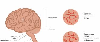
In addition, a violation of the strength of cell membranes at a reduced level provokes ruptures of the walls of blood vessels, which leads to hemorrhage in the brain - hemorrhagic stroke. The inability to absorb vitamins and minerals is accompanied by the occurrence of osteoporosis. In the elderly, the risk of dementia, emphysema, asthma, and COPD increases many times over.
Lowering cholesterol levels is especially dangerous due to the occurrence of psychogenic factors. These include prolonged depression, which occurs with addiction to alcohol or drugs, aggravated by suicidal tendencies.
Who is at risk for atherosclerosis
- Men over 35 years old.
- People with excess body weight (metabolic syndrome, obesity).
- Smokers.
- Patients with high blood pressure.
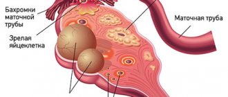
- Women with PCOS (polycystic ovary syndrome).
- Patients with type 2 diabetes.
- Patients whose close relatives have been diagnosed with atherosclerosis and cardiovascular diseases.
- Patients already suffering from atherosclerosis and cardiovascular diseases.
- Patients leading a sedentary lifestyle.
- Patients who abuse alcohol.
Evaluation of analysis results
When issuing the analysis result, the form will indicate the value obtained, as well as the boundaries to which to strive. These boundaries are not average intervals for a specific gender and age, as is usually used in reference values. When managing patients at risk of cardiovascular complications, doctors are primarily guided not by reference values, but by a decision-making threshold - target values of lipid profile indicators, according to the Recommendation of the third expert report of NCEP ATP III (The National Cholesterol Education Program Adult Treatment Panel III). These figures are calculated in accordance with population and clinical criteria, supported by numerous statistical data and approved by WHO.
Decision threshold criteria are very important diagnostic data, based on which the doctor, along with the patient’s test results and relying on his experience, knowledge and special risk calculation methods, can determine the most likely prognosis and select treatment for a particular patient.
The most well-known technique that helps a doctor determine the degree of risk for a patient is the so-called SCORE scale - Systematic COronary Risk Evaluation, which allows one to calculate the risk of death from cardiovascular diseases depending on the patient’s cholesterol level and blood pressure.
Hyperlipidemia syndrome in children and adolescents: pathogenesis, clinical picture, treatment
Lipids are organic substances that, together with proteins and carbohydrates, make up the bulk of the body's organic substances. They accumulate in adipose tissue in the form of triglycerides (TG), being the main energy substance. Lipids are part of cell membranes and participate in the synthesis of steroid hormones.
Physiology of lipid metabolism
Lipids without a protein connection are insoluble or poorly soluble in water, but as part of lipoprotein complexes consisting of lipids and proteins, they easily go into solution. In this state, they provide transport of lipids in the plasma for delivery of the latter to the body tissues.
The components of lipids are fatty acids (saturated, monounsaturated, polyunsaturated), triglycerides, free cholesterol (CS), cholesteryl esters (CE), phospholipids (PL, complex lipids).
TG are chemical compounds of glycerol with three molecules of fatty acids. In everyday life they are called fats. When TG is hydrolyzed in adipocytes in the presence of lipoprotein lipase (LPL), fatty acids are released from them, which are used by the body for energy purposes.
Cholesterol (chole - bile, sterol - fatty) is produced in the liver and is used for the construction of cell membranes, the synthesis of steroid hormones and bile acids. In the intestine, bile acids are necessary for the emulsification and absorption of exogenous fat.
The main form of cholesterol circulating in plasma is its esters (cholesterol bound to a fatty acid). They are in the form of highly soluble complexes with transporter proteins. Their formation requires the enzyme lecithin cholesterol acyltransferase (LCAT).
PL are lipids that contain glycerol, fatty acids, phosphoric acid, and nitrogen-containing compounds. They are an integral part of cell membranes.
In the blood, TG, cholesterol, and PL are transformed in the form of lipoproteins (LP), and free, non-fat, non-esterified fatty acids (NEFA) are transformed in the form of complexes with albumin [1].
In the process of ultracentrifugation, individual fractions of drugs are isolated: chylomicrons (CM) - the lightest particles, very low-density lipoproteins (VLDL), transition-density lipoproteins (TDLP), low-density lipoproteins (LDL), high-density lipoproteins (HDL).
The proteins that make up the drug are referred to as apolipoproteins (apo). They are necessary for the transport of various lipids (mainly triglycerides and cholesteryl esters) from sites of synthesis to the cells of peripheral tissues, as well as for the reverse transport of cholesterol to the liver. Apoproteins regulate the activity of enzymes - LCAT, LPL, hepatic triglyceride lipase (TGL).
There are six main classes of apolipoproteins, designated by letters (A, B, C, E, D, H). Their synthesis occurs mainly in the liver and intestines.
The main lipids of chylomicrons are TG, and their structural apoprotein is B48. VLDL consists of 55% TG, 20% cholesterol, and the transport protein is apo-B100.
DILI is formed from VLDL, contains 35% cholesterol and 25% TG and is transported by apo-B100. The LDLP fraction is an intermediate product, a precursor to LDL, which is formed during the conversion of VLDL with the participation of hepatic lipase. It is characterized by a short plasma lifetime and is rapidly metabolized into LDL.
LDL is the end product of VLDL hydrolysis. They are the main carriers of cholesterol to tissues. The half-life is 3–4 days. Approximately 70% of total cholesterol is associated with LDL. LDL catabolism occurs predominantly in peripheral tissues. In humans, 70–80% of LDL is removed from the plasma every day by receptor-mediated pathways. The rest is captured by phagocytic cells of the reticulohistiocytic system. It is believed that this is the main atherogenic lipoprotein. Apo-B100 makes up more than 95% of total LDL protein.
HDL is formed mainly in the liver, but also in the intestine from exogenous lipids. It is a heterogeneous group of molecules with distinct metabolic roles. HDL absorbs free PL cholesterol, which is formed in plasma under the influence of LCAT. High levels of HDL have a protective effect against atherosclerosis. HDL consists of 50% protein apo-A1, apo-E, apo-C, 25% PL, 20% cholesterol and 5% TG.
The formation of a lipid-protein complex occurs through many receptors. Changes in genes encoding apoprotein receptors can disrupt lipoprotein function [2].
A variety of fatty acids in the composition of chemicals are supplied with food, which are processed with the participation of hepatic lipase. The main lipids found in blood plasma are NEFA, PL, TG, cholesterol and EC. In a healthy person, CMs after consuming fats disappear from the blood 12 hours after eating.
An important source of fatty acid formation is carbohydrates. A significant portion of essential fatty acids is synthesized in the liver, less in adipocytes from acetyl-coenzyme A, formed during the conversion of glucose.
Insulin is a potent inhibitor of the activity of hormone-sensitive LPL and, consequently, lipolysis in the liver and adipose tissue. In adipocytes, high levels of glucose and insulin promote the deposition of FFAs in the form of TG. Insulin and glucose also stimulate the biosynthesis of FFAs in the liver when consumed in excess. FFAs are converted into TG with subsequent formation of VLDL. FFAs are captured by various cells and used as energy material, and when in excess they are deposited in the form of triglycerides in tissue.
Energy expenditure (lipolysis) occurs under the control of adrenaline, norepinephrine, glucagon, and adrenocorticotropic hormone (ACTH). The resulting FFAs capture working organs.
The content of cholesterol and TG in the blood plasma can be used to judge the nature of lipoprotein particles. An isolated increase in TG levels indicates an increase in the concentration of cholesterol or VLDL. On the other hand, an isolated increase in cholesterol levels almost always indicates an increase in LDL concentrations. TG and cholesterol levels often increase simultaneously. This may reflect a sharp increase in the concentration of cholesterol and VLDL [1, 2].
Hyperlipidemia
Plasma lipid levels vary among children due to genetic and dietary factors. Hyperlipoproteinemia (dyslipidemia) is a syndrome complex accompanied by excessive levels of lipids and/or lipids in the blood plasma.
Hyperlipidemia (HLP) occurs in 2–10% of children and 40–60% of adults [2]. HLP is an important factor in the development of cardiovascular diseases. LPs of one or more classes may accumulate in the blood due to increased formation or secretion or increased administration of exogenous lipid components or decreased excretion from the body. In some cases, all of these processes take place.
Disturbances in fat metabolism may be associated with changes in proteins involved in fat metabolism (LP, apoproteins) and in the LP receptor apparatus. The mechanism of these disorders may be due to a deficiency or block of apoproteins and LP receptors, which are the most important cofactors of LPL activity and endocytosis by macrophages. The synthesis of LDL receptors is inhibited by glucocorticoids.
GLP is divided into primary (1/3) and secondary (2/3). According to the mechanism of development, HLPs are grouped into nutritional, retention, and transport. In most cases they are combined.
According to the WHO classification, various combinations of drugs, the level of which is elevated in pathology, are divided into six types or categories. Most of them can be caused by various genetic diseases. Types of hyperlipoproteinemia should be considered as evidence of a disorder of lipid metabolism, and not as the name of a specific disease.
Primary SDP
Primary HLP type 1 (familial lipoprotein lipase deficiency) occurs in 1% of cases with autosomal recessive inheritance resulting from a mutation in the LPL gene. The defect is associated with congenital or acquired LPL deficiency. As a result of this disorder, the metabolism of CMs is blocked, which leads to their extreme accumulation in the plasma. With insufficient blood LPL activity, the transition of fatty acids from blood plasma chylomicrons to fat depots is disrupted (TGs are not broken down). The excretion of TG-rich drugs is blocked. Severe triglyceridemia develops.
The pathology usually manifests itself in early childhood with recurrent attacks of abdominal pain. They are caused by pancreatitis, which develops due to the formation of toxic products during the partial hydrolysis of TG and PL of chylomicrons. Excess CM can cause microthrombosis in various organs.
In the clinic, patients are found to have yellow papules with an erythematous rim in different areas of the skin and xanthoma as a result of the deposition of large quantities of chylomicron triglycerides in histiocytes. THs are also deposited in phagocytes of the reticulohistiocytic system, causing hepatomegaly, splenomegaly and bone marrow infiltration by foam cells. When examined with an ophthalmoscope, a whitish retina and white vessels in it can be detected, allowing the diagnosis of retinal lipemia. Despite the sharp increase in plasma TG levels, the development of atherosclerosis does not accelerate.
The diagnosis of familial lipoprotein lipase deficiency should be assumed when milky yellow, creamy blood plasma is detected upon standing. The diagnosis is confirmed by the absence of an increase in plasma lipoprotein lipase activity after heparin administration and a sharp increase in the levels of cholesterol and triglycerides.
Symptoms become less pronounced if the patient is switched to a low-fat diet. Since medium-chain TGs are not included in HM, these are the fats that should be used in the diet to ensure its normal calorie content. The patient must also receive fat-soluble vitamins.
HLP type 2 (familial hypercholesterolemia) is the most common form. It is an autosomal dominant disorder caused by a mutation in the gene for apo B100, a protein that binds LDL to the LDL receptor.
Due to the reduced activity of LDL receptors, the catabolism of these drugs is blocked, and their amount in the plasma increases in proportion to the decrease in receptor function. The liver is unable to effectively capture LDLP, which should be converted into LDL. This leads to impaired uptake, impaired LDL excretion and accumulation of total cholesterol in the blood plasma. As a result, their concentration increases by 2–6 times.
This form of HLP is divided into subtypes 2a and 2b. The first is characterized, along with an increase in LDL and cholesterol, by normal levels of VLDL and TG. With subtype 2b, described in 1973, combined lipidemia is typical: increased LDL, VLDL, TG, cholesterol.
Heterozygous familial hypercholesterolemia type 2a should be assumed when an isolated increase in plasma cholesterol levels is detected in children against the background of unchanged triglyceride concentrations. An isolated increase in cholesterol levels is usually caused by an increase in the concentration of LDL cholesterol only. In familial hypercholesterolemia, only half of first-degree relatives have elevated plasma cholesterol levels. The presence of skin xanthomas helps in diagnosis. When collecting anamnesis from relatives, early manifestations of coronary heart disease can be identified.
HLP type 2b is much more common in children. High levels of cholesterol, TG, LDL, and VLDL are detected. Excess LDL infiltrates the vascular wall, leads to unregulated accumulation of cholesterol in cells, their foamy transformation, and contributes to the development and early progression of atherosclerosis and coronary heart disease.
Relatives are often diagnosed with hyperinsulinism, insulin resistance, arterial hypertension, uricemia, and early development of atherosclerosis. With familial hypercholesterolemia, the incidence of obesity and diabetes mellitus does not increase. In patients, body weight, as a rule, is even less than normal.
This form of HLP occurs already in newborns. With homozygous carriage, such patients die in childhood from myocardial infarction. In cases where very high levels of cholesterol in the blood plasma are detected, parents should be examined to exclude familial hypercholesterolemia.
Since atherosclerosis in this disease is caused by a prolonged increase in plasma LDL levels, patients should be switched to a diet low in cholesterol and saturated fat and high in polyunsaturated fat. You should exclude butter, cheese, chocolate, fatty meats and add polyunsaturated oils (corn, olive, fish oil, etc.). In this case, the plasma cholesterol level in heterozygotes decreases by 10–15% [1].
Familial HLP type 3 is a rare form. In this congenital disease, plasma levels of both cholesterol and TG are elevated. This is due to the accumulation of residues in the plasma resulting from the partial destruction of VLDL. The mutation that determines the disease affects the gene encoding the structure of apoprotein E, a protein that is normally found in DILI and remnants of chylomicrons. It binds both the chylomicron remnant receptor and the DILI receptor with very high affinity. VLDL, LPPP, and TG increase in the blood plasma. The conversion of VLDL to LDL is disrupted, and the level of HDL decreases. These drugs accumulate in large quantities in plasma and tissues, causing xanthomatosis and atherosclerosis.
HLP manifests itself after 20 years. In the clinic, accelerated sclerosis of the vessels of the heart, carotid arteries, and vessels of the lower extremities is typical, and, consequently, early myocardial infarctions, strokes, intermittent claudication and gangrene of the legs.
The disease may be accompanied by obesity, type 2 diabetes mellitus, and hepatosis. Tuberous xanthomas, which are localized in the area of the elbow and knee joints, are considered pathognomonic. The plasma becomes cloudy when standing.
Primary type 4 HLP, or familial triglyceridemia, is characterized by high TG concentrations with normal or slightly elevated fasting cholesterol levels. It is a common (17–37%) autosomal dominant disorder. It appears in various periods of childhood, but more often in older years.
The pathogenesis of primary triglyceridemia is not entirely clear. It is believed that there is excess production of TG in the presence of normal levels of apo-B lipoproteins, causing the formation of TG-rich VLDL in the liver. There is not enough data on the atherogenicity of these lipoproteins [2].
Familial triglyceridemia can be suspected if the patient has high levels of TG, VLDL and a normal amount of cholesterol. The disease should be confirmed by studying the lipid profile of close relatives. When the blood taken is left standing for 8 hours, flakes can be detected in the plasma. In some patients, HDL is elevated, which may be due to the low atherogenicity of this form of HLP. Xanthomas are not typical for familial hypertriglyceridemia.
This variant of secondary HLP occurs in metabolic syndrome, type 2 diabetes mellitus and other endocrinopathies.
HLP type 5, or familial combined HLP, is inherited as an autosomal dominant trait. This form, combining an increase in TG and CM, has signs of types 1 and 4 of HLP. Close relatives may have only a mild form of the disease with moderate hypertriglyceridemia without hyperchylomicronemia, hypercholesterolemia, i.e., it may be characterized by hyperlipidemia of various types. When standing, the plasma forms a cloudy layer. The mutant gene is unknown.
Increases in plasma cholesterol and/or triglyceride levels are detected at puberty and persist throughout the patient's life. The increase in lipid and lipid levels is not constant: during different periods of the examination, high indicators may decrease, while others, on the contrary, may increase significantly.
Family history often suggests early coronary artery disease. Mixed type HLP is found in approximately 10% of all patients with myocardial infarction. The incidence of obesity, hyperuricemia and impaired glucose tolerance is increased [1].
Mild or moderate hypertriglyceridemia can sharply increase under the influence of various provoking factors (decompensated diabetes mellitus, hypothyroidism, taking medications containing estrogens). Plasma triglyceride levels may increase. During periods of exacerbations, patients develop mixed hyperlipidemia, i.e., the concentration of both VLDL and CM increases. After eliminating the provoking factors, chylomicron-like particles disappear from the plasma and the concentration of triglycerides returns to the original level.
Secondary SLP
There are basal and induced HLP. Basal HLP is detected in fasting blood tests and is more severe and indicates profound disturbances in fat metabolism. Induced (postprandial) HLP are detected after taking carbohydrates, fats, hormonal drugs, and severe physical activity. These include nutritional HF, which develop after excessive consumption of fats in food.
Secondary HLP are often manifested by increased levels of several classes of drugs in the blood plasma. The change in lipid spectrum may be similar to the primary types of HLP. Clinical changes are determined by the underlying disease. Often (primarily at the beginning of the development of the pathological process), quantitative and qualitative changes in the composition of drugs in the blood plasma are adaptive in nature [3].
Diabetes mellitus type 1
Insulin-dependent diabetes mellitus (DM) significantly affects lipid metabolism. Hypertriglyceridemia occurs in most patients. It is associated with the role of insulin in the formation and removal of lipid-rich drugs from plasma [1, 3].
With insulin deficiency, the synthesis of TG and the content of apo-B lipoproteins increase. Due to impaired LPL activity, the excretion of TG-rich particles is reduced. Insulin deficiency increases lipolysis in adipose tissue, resulting in a high flow of FFA to the liver. It enhances the synthesis and secretion of TG into the blood as part of VLDL as a secondary reaction to the increased mobilization of free fatty acids from adipose tissue.
The cholesterol content of LDL increases, but to a lesser extent. In decompensated diabetes, the plasma concentration of FFA is increased and correlates with the level of glucose in the blood.
As the duration of hypoinsulinemia increases, the rate of removal of VLDL and cholesterol from the blood decreases due to a decrease in lipoprotein lipase activity, since insulin is necessary for the synthesis of LPL in adipocytes. Diabetic lipimia develops. This is facilitated by glucagon, which increases sharply during decompensation, increasing the release of FFA into the blood.
Some of the FFA is metabolized to acetyl-CoA, which is then converted into acetoacetic and B-hydroxybutyric acids. These lipid metabolism disorders resemble the phenotype characteristic of primary type 4 or 5 HLP.
Severe lipidemia leads to fatty liver and pancreas, creamy appearance of retinal vessels, and abdominal pain.
According to V.M. Delyagin et al. In children with type 1 diabetes, dyslipidemic disorders were manifested by changes in the pattern of the liver parenchyma, thickening of the vascular walls, changes in the media/intima ratio, and a decrease in vascular elasticity [4].
The blood has a milky color and a creamy layer appears when standing. This is due to acquired LPL deficiency. But, given the prevalence of diabetes, a combination of primary hereditary disorders of lipid metabolism and HLP associated with absolute insulin deficiency is possible. Hormone replacement therapy quickly reduces the severity of lipoproteinemia.
So, hypertriglyceridemia, increased VLDL cholesterol levels, and non-enzymatic glycosylation of apoproteins are risk factors for the development of atherosclerosis in patients with type 1 diabetes and type 2 diabetes. Compensation of diabetes reduces the severity of HLP and, therefore, inhibits the development of atherosclerosis.
Obesity
Among all forms of obesity in childhood, a simple form occurs (85–90%). The constitutional factor is leading in the genesis of this form of the disease. Genetic predisposition to obesity may be associated with central dysregulation of energy balance, with an increase in the number of adipocytes and increased metabolic activity of the latter.
In adipose tissue, the synthesis and hydrolysis of lipids, the synthesis of fatty acids, including from carbohydrates, their esterification into neutral fats (TG), their deposition and breakdown with the formation of fatty acids, and their use for energy purposes constantly occur.
LPL activity in adipose tissue is regulated by insulin, i.e. insulin stimulates the uptake and accumulation of circulating FFAs in adipocytes.
Another mechanism for lipid deposition in adipose tissue involves the conversion of glucose and other carbohydrates into fats, which is controlled by insulin. Under physiological conditions, one third of dietary glucose is used for the synthesis of endogenous fat, in obese patients up to two thirds.
In the blood of obese patients, increased levels of cholesterol, TG, LDL and decreased HDL are detected. Such patients have reduced tolerance to exogenous lipids—in response to a fat load, they experience a pronounced prolonged hyperlipidemic reaction. CMs in these patients are broken down slowly, and lipolytic activity appears late [3].
Hyperinsulinemia is important in the pathogenesis of obesity. Increased insulin secretion causes increased appetite. The liver begins to synthesize more TG.
Metabolic syndrome
Obesity with excess visceral fat, accompanied by insulin resistance (IR) and hyperinsulinemia (HI), disorders of carbohydrate, lipid, purine metabolism, and arterial hypertension, is systematized under the term “metabolic syndrome (MS)” [5].
Abdominal obesity increases the release of FFAs from adipocytes into the blood plasma and their uptake by the liver. The synthesis of TG and apo-B lipoproteins increases in it, and small LDL particles dominate. The production of HDL decreases, the removal of excess cholesterol from the body is impaired, the level of glucose in the blood increases, and the tendency to blood clots and inflammation increases. Conditions are created for the formation of the atherosclerotic process. This is predisposed by glycation of apoproteins, which impairs their binding to lipids. Glycated VLDL cholesterol and LDL cholesterol circulate in the blood longer. The risk of developing cardiovascular diseases increases.
The pathogenetic cause of MS is still unknown, but a close relationship has been noted between increased body weight and IR and GI. Abdominal obesity contributes to increased IR, causing an increased influx of FFA from adipocytes to the liver. Adipocytes of visceral adipose tissue, having increased sensitivity to the lipolytic action of catecholamines and low sensitivity to the antilipidic action of insulin, secrete FFAs, which prevent the binding of insulin to the hepatocyte, which leads to HI [6].
Risk factors for metabolic syndrome in a child are the presence of abdominal obesity, type 2 diabetes, arterial hypertension, myocardial infarction, stroke and other vascular disorders in relatives [2].
To diagnose MS, it is necessary to evaluate anthropometric measurements, blood pressure, determination of glucose, insulin, fasting C-peptide levels, and lipid spectrum. A simple indicator is the IR ratio of fasting TG concentration to HDL cholesterol level. If this index is 3.5 or more, then this indicates IR.
Hypothyroidism
Dyslipidemia accompanying hypothyroidism is accompanied by an increase in atherogenic drugs and a decrease in the content of antiatherogenic drugs.
Thyroid hormones influence all stages of lipid metabolism. They promote the uptake and synthesis of lipids in the liver, the oxidation of TG from adipose tissue, the transport of FFAs, the absorption and excretion of cholesterol in the composition of bile acids, and an increase in the activity of hepatic lipase.
Apparently, thyroid hormones exert their effects by stimulating genes responsible for the synthesis and lipolysis of lipids and lipids. With a deficiency of thyroid hormones, the amount of apo-B and apo-E proteins and LDL receptors in the liver decreases, the excretion of cholesterol is impaired, and the level of VLDL and LDL in the blood increases. A decrease in hepatic lipase activity disrupts the conversion of VLDL into LDLP, and from the latter the formation of HDL. A decrease in the activity of cholesterol transport protein leads to disruption of the reverse transport of cholesterol to the liver.
A high level of LDL cholesterol with a reduced concentration of HDL is associated with a high risk of atherosclerosis [7].
If hypothyroidism is detected, levothyroxine is prescribed. This treatment is accompanied by normalization of the patient's lipid levels within a month.
In subclinical hypothyroidism, the presence of atherogenic dyslipidemia is also indicated, which requires corrective therapy. Opinions are divided on thyroid hormone replacement therapy for subclinical hypothyroidism [7, 8]. Some authors believe that the administration of L-thyroxine more often normalizes the lipid spectrum in such patients. Others believe that the administration of lipid-lowering drugs is sufficient to correct dyslipidemia.
Somatotropic insufficiency
Growth hormone (GH) plays an active role in the regulation of basal metabolism. It is an anabolic hormone and has lipolytic activity. In adipose tissue, GH increases the number of preadipocytes and suppresses the activity of LPL, an enzyme that hydrolyzes TG in lipoproteins to NEFA, which are re-esterified in the liver and deposited in adipocytes [9].
Most patients with somatotropic insufficiency (STI) exhibit excess adipose tissue, decreased muscle mass and strength, osteoporosis, and early atherosclerosis. GH deficiency in adults increases the risk of death from cardiovascular diseases (myocardial infarction, stroke, etc.). One of the factors that provoke changes in blood vessels during STN is a disorder of lipid metabolism. An increase in the level of total cholesterol, LDL, and apoprotein B is detected. Other studies have revealed an increase in TG levels and a decrease in HDL [10]. More significant changes in lipid metabolism were observed in patients with multiple pituitary hormone deficiency.
Long-term replacement therapy with recombinant growth hormone led to a decrease in the levels of total cholesterol and LDL, but did not affect the content of HDL and TG [11].
Cushing's syndrome
Glucocorticoids (GCs) play important roles in energy homeostasis and have complex but unclear effects on lipid metabolism. GCs modulate the expression of about 10% of human genes. They regulate the differentiation, function and distribution of adipose tissue.
Under the influence of GCs, FFAs in the blood increase, since these hormones accelerate lipolysis, disrupt the formation of glycerol, and reduce glucose utilization. The effect of GC on fat metabolism depends on the localization of subcutaneous fat. With an excess of HA, the fat layer on the extremities is reduced, and on the face and torso it is overdeveloped.
With an excess of glucocorticoids in the blood, total cholesterol and TG increase, while the level of HDL may vary. Insulin resistance plays a key role in this dyslipidemia. In vitro, cortisol increases lipoprotein lipase levels in adipose tissue, especially visceral fat, where lipolysis is activated and FFAs are released into the circulation.
Impaired glucose tolerance or steroid diabetes, IR, hypercoagulation determine an increased risk of cardiovascular pathology in this group of patients. However, the degree of dyslipidemia in the clinic is very variable in patients with exogenous and endogenous excess GC [1, 2].
A study of 15,004 patients found that GC use was not directly associated with an unfavorable lipid profile. In Cushing's syndrome, there is an increase in VLDL, but not HDL, with a subsequent increase in triglycerides and total cholesterol. The prevalence of hepatic steatosis among patients receiving GCs and patients with Cushing's syndrome is not the same. In the latter, it primarily depends on the volume of abdominal and visceral fat [12].
These changes are normalized or significantly improved after correction of hypercortisolism.
Kidney diseases
HLP in renal diseases is common. Changes in the spectrum of lipids occur already in the early stages of glomerulonephritis and are aggravated during chronic course. In the acute period of the disease, an increase in TG, represented by cholesterol and VLDL, is characteristic, i.e., dyslipidemia develops, similar to primary type 4 HLP. The cholesterol content in VLDL also increases with a relative decrease in TG. Opposite ratios are typical in LDL.
In chronic kidney disease, dyslipidemia is manifested by an increase in TG. This is due to an increase in the content and decrease in clearance of VLDL and accumulation of LDL [13]. The experiment showed that in case of renal failure, the formation of receptors for VLDL in adipose tissue is disrupted, which increases their concentration and, consequently, maintains hypertriglyceridemia. This is facilitated by a decrease in LPL activity in adipose tissue. Deficiency of hepatic lipase impairs the hydrolysis of TG in LDLP with the formation of LDL and HDL phospholipids.
With renal failure, the content of the most atherogenic LDPP increases, which accelerates the atherosclerotic process.
With nephritic syndrome, a mixed form of HF is often detected with an increase in both cholesterol and TG, which is similar to type 2 of HF. The concentration of LDL increases with normal or decreased amount of HDL. HLP is associated with hypoalbuminemia. Remission of the disease is accompanied by normalization of lipidemia.
Treatment
Etiological treatment for primary HLP has not yet been developed. Some forms of secondary HLP are well corrected by replacement therapy (type 1 diabetes, hypothyroidism, somatotropic insufficiency).
Pathogenetic treatment of HF is primarily aimed at reducing plasma cholesterol levels. A diet low in cholesterol and saturated fat and high in polyunsaturated fat. Exclude butter, cheese, chocolate, pork. The diet uses corn and sunflower oil.
When metabolic syndrome is detected, it is recommended to limit or eliminate easily digestible carbohydrates. The amount of fat is reduced by up to 70%, mainly due to animals, which are replaced by 50% with vegetable ones.
Physical activity along with diet for HLP is the most important factor in treatment. Physical activity increases metabolism and accelerates the mobilization of fat from the depot.
The use of lipid-lowering drugs in childhood is limited due to many side effects [14].
Literature
- Kronenberg G. M. et al. Obesity and lipid metabolism disorders / Trans. from English edited by I. I. Dedova, G. F. Melnichenko. M.: Read Elsiver LLC, 2010. 264 p.
- Endocrinology and metabolism. T. 2. Per. from English / Under. ed. F. Fehling, J. Baxter, A. E. Broadus, L. A. Fromen. M.: Med., 1985. 416 p.
- Turkina T.I., Shcherbakova V.Yu. Features of dyslipidemia in children // Rational pharmacotherapy in cardiology. 2011, no. 7 (1), 65–69.
- Delyagin V.M., Melnikova M.B., Serik G.I. Dyslipidemia syndrome in children with chronic diseases // Practical Medicine. 2014, No. 9 (85), p. 7–10.
- Bokova T. A. Metabolic syndrome in children. Tutorial. M., 2013. 21 p.
- Gurina A. E., Mikaelyan N. P., Kulaeva et al. The relationship between the activity of insulin receptors and blood ATP against the background of dyslipidemia in children with diabetes mellitus // Fundamental Research. 2013, no. 12–1, p. 30–34.
- Biondi B., Klein I. Cardiovascular abnormalities in subclinical and overt hypothyroidism/The Thyroid and cardiovascular risk. Stuttgart; New York, 2005. 30–35.
- Budievsky A.V., Kravchenko A.Ya., Feskova A.A., Drobysheva E.S. Dyslipidemia in subclinical hypofunction of the thyroid gland and the effectiveness of its correction with L-thyroxine replacement therapy // Young scientist. 2014, 17, p. 138–141.
- Dedov I. I., Tyulpakov A. N., Peterkova V. A. Somatotropic insufficiency. M.: Index Print, 1998. 312 p.
- Volevodz N. N. Systemic and metabolic effects of growth hormone in children with various types of short stature. Diss. Doctor of Medical Sciences 2005.
- Volevodz N. N., Shiryaeva T. Yu., Nagaeva E. V., Peterkova V. A. The state of the lipid profile in patients with somatotropic insufficiency and the effectiveness of correction of dyslipidemia during treatment with the domestic recombinant growth hormone “Rastan”. https://umedp.ru |articles|sostoyanie_lipidnogo_profilya.
- Arnaldi G., Scandali VM, Trementino L., Cardinaletti M., Appolloni G., Boscaro M. Pathophysiology of Dyslipidemia in Cushing's Syndrome // Neuroendocrinology. 2010; 92(suppl 1): 86–90.
- Rudenko T. E., Kutyrina I. M., Shvetsov M. Yu. State of lipid metabolism in chronic kidney disease. Clinical nephrology. 2012, no. 2, p. 14–21.
- Vasyukova O. V. Federal clinical guidelines for the diagnosis and treatment of obesity in children and adolescents. M.: Institute of Pediatric Endocrinology ENC, 2013. 18 p.
V. V. Smirnov1, Doctor of Medical Sciences, Professor A. A. Nakula
State Budgetary Educational Institution of Higher Professional Education Russian National Research Medical University named after N. I. Pirogov Ministry of Health of the Russian Federation, Moscow
1 Contact information
Decision thresholds
For total cholesterol , according to experts from NCEP ATP III (The National Cholesterol Education Program Adult Treatment Panel III), the optimal value should be below 5.2 mmol/l. This is the average upper limit to which we must strive. Values in the range of 5.2–6.1 mmol/l are considered borderline. With values above 6.1 mmol/l, we can talk about serious risks of developing atherosclerosis.
For LDL, the optimal value is no more than 2.6 mmol/l. Values from 2.6 to 3.3 mmol/l are considered borderline. Above 3.3 mmol/l - the risk of developing atherosclerosis.
normal HDL level For patients at risk, as well as for patients already suffering from atherosclerosis, the target value is higher - more than 1.55 mmol/l.
For triglycerides, a value of up to 1.7 mmol/l is considered normal. From 1.7 to 2.2 mmol/l - borderline value, above 2.2 mmol/l - dangerously elevated, associated with the risk of developing atherosclerosis. However, it should be remembered that an increase in triglycerides in itself may not be a sign of atherosclerosis or lipid metabolism disorders. So, this may be due to the fact that the patient did not properly prepare for the test (could not withstand a 10-12 hour fast) or other pathological processes.
An important indicator of the risk of developing atherosclerosis is the atherogenic coefficient (AC) .
The formula for calculating it is as follows:
The normal KA level for young people 20–30 years old is up to 2.7. For people over 30 years of age, the value can be up to 3–3.5.
A KA indicator of 4 or higher is a sign of the onset of atherosclerosis and the risk of developing cardiovascular diseases. In this case, your doctor will usually prescribe medication to lower your cholesterol levels.
Cholesterol-VLDL (calculated value: total cholesterol, HDL, LDL, triglycerides)
Cholesterol is a fat-like substance vital to the body. With its help, the formation of cell membranes of all organs and tissues of the body occurs. Hormones are created on the basis of cholesterol, which are involved in the growth, development of the body and the implementation of the reproduction function. In addition, bile acids, which are part of bile, are formed from cholesterol, thanks to which fats are absorbed in the intestines. Cholesterol is insoluble in water, so in order to move around the body it is “packed” into a shell consisting of special proteins - apolipoproteins. The resulting complex (cholesterol + apolipoprotein) is called lipoprotein. Several types of lipoproteins circulate in the blood, differing in the proportions of their components:
- very low density lipoproteins (VLDL),
- low density lipoproteins (LDL),
- high density lipoproteins (HDL).
Very low-density lipoprotein cholesterol is one of the most aggressive types of cholesterol. When there is an excess of VLDL cholesterol, it is deposited on the walls of blood vessels in the form of plaques, which can restrict the movement of blood through the vessel. In addition, they make blood vessels more rigid (atherosclerosis), which significantly increases the risk of heart disease (coronary artery disease, heart attack) and stroke. In addition, VLDL is the main carrier in the body of another type of fat - triglycerides. Elevated triglyceride levels also contribute to the development of atherosclerosis. The liver produces enough cholesterol and triglycerides for the body's needs, but some of these fats come from food, mainly meat and dairy products. If a person has a family history of high cholesterol and triglycerides or eats too much cholesterol-containing food, cholesterol levels in the blood can rise and cause harm to the body. What is analysis used for? A VLDL cholesterol test, along with other tests included in the lipid profile, is necessary in order to assess the risk of developing atherosclerosis and heart problems. Atherosclerosis is the process of plaque growth inside blood vessels, which can limit blood flow through the vessel or completely block its lumen. In addition, this study can be conducted to monitor the effectiveness of a diet with a reduced amount of animal fats and monitor blood lipid levels after the prescription of cholesterol-lowering drugs. When is the test scheduled? VLDL is usually included in the lipid profile, along with the determination of total cholesterol, LDL cholesterol, HDL cholesterol, triglycerides and atherogenicity coefficient. A lipidogram can be prescribed during routine preventive examinations or when total cholesterol is increased in order to find out what type of cholesterol is increasing. A lipidogram is recommended for all adults over 20 at least once every 5 years. It may be prescribed more often (several times a year) if a person is prescribed a diet limiting animal fats and/or is taking medications that lower cholesterol levels. In these cases, it is checked whether the patient has achieved the target level of lipid values and, accordingly, whether his risk of cardiovascular diseases is reduced.
Increased levels of VLDL cholesterol Increased concentrations of VLDL cholesterol may be the result of a hereditary predisposition (familial hypercholesterolemia) or excess dietary intake of animal fats. For most people with high cholesterol, both causes are involved to some extent. Other possible causes of increased VLDL:
- cholestasis - stagnation of bile, which can be caused by liver disease (hepatitis, cirrhosis) or gallstones,
- chronic inflammation of the kidneys leading to nephrotic syndrome,
- chronic renal failure,
- decreased thyroid function (hypothyroidism),
- poorly treated diabetes mellitus,
- alcoholism,
- obesity,
- prostate or pancreatic cancer.
Decrease in VLDL cholesterol levels
- A reduced level of LDL cholesterol has no particular clinical significance; it can be observed in the following conditions:
- hereditary hypocholesterolemia,
- severe liver disease,
- oncological diseases of the bone marrow,
- increased thyroid function (hyperthyroidism),
- inflammatory joint diseases,
- B12 or folate deficiency anemia,
- common burns,
- acute diseases, acute infections,
- chronic obstructive pulmonary disease.
What can influence the result? The amount of VLDL cholesterol can change from time to time, so a one-time measurement does not always reflect the “usual” cholesterol level. In this regard, it is sometimes necessary to re-take the test after 1-3 months. It happens that the level of VLDL cholesterol increases or decreases over a short period of time. This phenomenon is called biological variation and reflects normal fluctuations in cholesterol metabolism in the body. Increases VLDL cholesterol levels:
- pregnancy (lipid profile should be done at least 6 weeks after birth),
- long fasting,
- donate blood while standing,
- anabolic steroids, androgens, corticosteroids,
- smoking,
- eating food containing animal fats.
Reduce VLDL cholesterol levels:
- being in a lying position,
- allopurinol, clofibrate, colchicine, antifungal drugs, statins, cholestyramine, erythromycin, estrogens,
- intense physical activity,
- a diet low in cholesterol and saturated fatty acids and, conversely, high in polyunsaturated fatty acids.
The following contribute to an increased risk of developing cardiovascular diseases:
- smoking,
- age (men over 45 years old, women over 55 years old)
- increased blood pressure (140/90 mmHg and above),
- increased cholesterol or cardiovascular disease in other family members (heart attack or stroke in the closest male relative under 55 years of age, female under 65),
- coronary heart disease, previous myocardial infarction or stroke,
- diabetes,
- excess body weight,
- alcohol abuse,
- eating large amounts of food containing animal fats,
- low physical activity.
How you can improve your lipid profile
Power change:
- Choose foods that are low in total fat: Keep fat in your diet to no more than 30% of total calories.
- Choose foods that contain Omega-3 unsaturated fatty acids. Omega-3s are found in fatty fish (salmon, tuna, sardines, herring, mackerel and other cold-water fish), and in smaller quantities in flaxseed oil, soybeans, spinach, and broccoli. However, it should be remembered that people suffering from cardiovascular diseases should avoid salt, so fresh fish is preferable to salted fish. Dietary supplements high in Omega-3 can also be a convenient and safe source of Omega-3.
- Limit the amount of red meat in your diet.
- Choose low-fat dairy products: one percent or skim milk, cottage cheese or yogurt.
- Choose foods high in complex carbohydrates and fiber: whole grain breads and cereals, fresh fruits, leafy vegetables, beans and lentils.
- Opt for plant-based proteins: Eat lentils, soy and tofu, with beans and legumes as alternatives, 2-3 times a week or more.
Other lifestyle changes:
- Maintain a healthy weight: Talk to your healthcare provider about your body mass index (BMI). BMI is measured by the ratio of your height to your weight.
- Exercise regularly for at least 20-40 minutes 3-4 times a week.
- Stop smoking. Smoking is an important additional risk factor that aggravates the course of atherosclerosis and negatively affects the lipid profile.
Is low cholesterol good or bad?
The most famous of all fatty compounds in the body is, of course, cholesterol. Quite a lot is said and written about this substance and its effect on the body. But usually they mention an increased level of total cholesterol or its “bad” fraction, which forms damage to blood vessels and fatty plaques on their walls. This condition is called atherosclerosis, it is dangerous because damaged vessels cope worse with their function - delivering blood to organs and tissues. High cholesterol levels are also associated with an increased risk of diabetes, hypertension, heart attacks and strokes.

Is it good to have low cholesterol?
Much has been written about how you need to adjust your diet to lower cholesterol levels, suggesting ways to keep it under control through physical activity and taking medications. However, there are situations when the opposite condition occurs - low cholesterol. It would be logical to conclude that if high cholesterol is bad, then very low cholesterol is good. But the body is a balanced system; sharp deviations in indicators, both downward and upward, are dangerous for it. Therefore, low cholesterol levels can also be dangerous.
Although high levels of total and “bad” cholesterol are dangerous for human health, and it is necessary to correct its concentration in the blood by all means, it is impossible to achieve a significant reduction in it. Recent studies have found that extremely low levels of cholesterol are just as dangerous. But even doctors rarely pay attention to lowering cholesterol, so there is very little data on how to eliminate or treat hypocholesterolemia.
The role of cholesterol in the body
Cholesterol is a substance that is vital for the body. Although an increase in the level of this compound in the blood can be a signal of the onset of various health problems, it is important to understand that cholesterol itself is not harmful, and you should not try to reduce its level if the levels are completely normal. Low cholesterol levels, according to scientists, are no less dangerous than high ones. To understand why cholesterol levels should be constant, you need to know why the body needs these fat molecules.
First of all, cholesterol is an integral part of cell walls, it helps create membrane strength and protects cells from damage. Equally important is a sufficient amount of cholesterol so that the body can produce steroid (including sex) hormones. In addition, the liver synthesizes bile from cholesterol molecules, a thick yellow liquid that helps break down and digest fatty foods. In addition, cholesterol helps transform inactive forms of vitamin D into active forms in the skin. Due to a sufficient amount of cholesterol, other fat-soluble vitamins are actively absorbed - these are A, E and K.
Why might cholesterol decrease?
There is evidence that low cholesterol levels are possible in people who play sports professionally. This is due to the fact that they rationally and actively spend fat accumulations and receive enough energy for physical activity. Their cholesterol does not accumulate, but is immediately used for the needs of the body. In addition, there is a small percentage of people who, despite being completely healthy, have slightly lower cholesterol levels when tested in a blood test. If the deviations are sharp, exceeding 10% of the standard values, this should be a reason for a more targeted examination. Only 20% of cholesterol comes from food, the remaining 80% is produced by the body itself. Therefore, low levels of cholesterol compounds may be a signal of metabolic problems. In rare cases, the defect is hereditary in nature; all family members have low cholesterol levels, but if this is detected in a relatively healthy person, it is important to know what factors can cause it.

Very low plasma cholesterol levels are possible with:
Liver pathologies.
It is the liver that plays a leading role in the metabolism of cholesterol and many other fatty substances. Therefore, any problems with the organ, especially if its cells become inflamed or damaged, leads to changes in fat levels. Key diseases include penile tumors, viral hepatitis and serious endocrine-metabolic lesions (fatty hepatosis, alcoholic lesions, metabolic syndrome). Against the background of a serious decrease in the activity of liver cells, cholesterol levels decrease - the liver simply cannot synthesize enough of it.
Pathologies of the thyroid gland.
A sharp decrease or excessive activity of the organ, called hypothyroidism or thyrotoxicosis, respectively, provokes cholesterol metabolism disorders. Although it is not completely clear how the thyroid gland affects metabolic processes, including fat metabolism, against the background of thyrotoxicosis, when the thyroid gland synthesizes too much thyroxine (T4 hormone), cholesterol levels sharply decrease. Along with this, weight decreases, body temperature rises, arrhythmias, sweating, memory and attention problems, and body tremors occur.
Intestinal absorption disorders.
There are a number of intestinal problems that interfere with the complete absorption of food components. An example is celiac disease (gluten enteropathy), which may impair the absorption of fats, including cholesterol. If the functioning of intestinal enzymes is disrupted, malabsorption syndrome occurs (impaired intestinal absorption), and cholesterol levels decrease. Regardless of what and in what quantities the patient eats, in case of serious intestinal problems, cholesterol will decrease.
Systematic fasting and diets for weight loss.
Often, due to malnutrition and constant weight loss, the supply of nutrients and minerals decreases. Particularly affected is the amount of manganese, which is associated with lower cholesterol levels. This fact is still being studied, but in laboratory conditions the connection between low cholesterol and a deficiency of manganese and other minerals has already been proven.
Why low cholesterol is dangerous: neurological disorders
Not so long ago, the fact was made public that a pathological decrease in cholesterol levels in both men and women is associated with mental disorders. This is especially true with regard to the development of pathological anxiety and depression. In addition, researchers from South Korea found that among patients prone to suicide, many more people had low cholesterol.
Hypocholesterolemia is believed to be associated with behavioral disorders such as aggression and impulsivity. There is also evidence that people who take high doses of cholesterol-lowering drugs are more likely to die from pathologies and causes not related to the heart and blood vessels. Often these are disasters and accidents, accidents and various problems associated with impulsive behavior.
But the most dangerous thing, which is being especially closely studied, is the risk of hemorrhagic stroke (or cerebral hemorrhage). There is evidence that very low cholesterol increases the risk of this complication by 5 times.

Digestive problems and cholesterol reduction
Low cholesterol is typical for thin people, patients who suffer from digestive problems or have poor nutrition, and are constantly on a diet. Impaired absorption of cholesterol from food and metabolic problems may partly explain hypocholesterolemia. There is a category of people who can eat a lot of any food without gaining weight, and they also often have lower cholesterol levels. a detailed examination may reveal gastritis, pancreatitis or biliary dyskinesia. Treatment helps you gain weight and normalize digestion, but cholesterol may remain low. In this case, they are advised to consume more saturated, animal fats. Although this goes against the principles of a healthy diet, it is for these people that such a diet can be a salvation from possible problems. If cholesterol is not corrected, there may be breathing problems, a tendency to bleed heavily, heart problems and depression, difficulty concentrating and accidents.
For expectant mothers
Research conducted several years ago at the Human Genome Research Institute (USA) found that low cholesterol during pregnancy can increase the risk of premature birth. In addition, scientists also found that hypocholesterolemia in the mother can negatively affect the development of the fetus, causing respiratory disorders and decreased birth weight.
Association with serious illnesses
You need to understand that none of the body’s compounds decreases without real reasons. Therefore, low cholesterol is a serious problem, the significance of which is still being studied. There are publications linking lower cholesterol levels with cancer (myeloma, leukemia, metastatic cancer). There is also evidence that hypocholesterolemia is associated with chronic infections, autoimmune diseases, premature aging and the provocation of systemic inflammation. All these conditions negatively affect human health.

Dietary fats and cholesterol: should they be reduced?
Naturally, constantly hearing about the dangers of cholesterol, people, willy-nilly, begin to be prejudiced against all saturated fats. They try to completely eliminate foods with animal fat and cholesterol from their diet. But this is wrong, the body is designed to receive all possible components from food, including saturated animal fat. The only exceptions are artificial, hydrogenated, and trans fats. They definitely will not benefit the body.
If we are talking about animal fat of natural origin (milk, animal meat), small amounts of these substances are needed by the body.
Firstly, it is a good source of energy for the body, a reserve fuel in case of starvation.
Secondly, fats create a protective layer around organs under the skin, protecting against concussions, blows and hypothermia.
Problems with fat metabolism usually arise with excess consumption of carbohydrates and fats, especially unnatural ones (hydrogenated, trans fats), against the background of a deficiency of protein and omega-3 acids. In addition, a person consumes a lot of hidden fats with industrially produced products, which are added to reduce the cost of products and increase their taste (but not usefulness).