Computed tomography of the adrenal glands is performed by scanning tissues and organs with X-rays. During the study, layer-by-layer images of organ projections are projected on the computer part of the tomograph in the form of a three-dimensional model. They reflect with high anatomical accuracy the state of all structures of this endocrine gland.
Sometimes, to maximize tissue contrast, a CT scan of the adrenal glands with a contrast agent is used. Contrast makes it possible to better identify neoplasms and inflammatory pathological changes in the body at the stage of their inception. The administration of contrast by intravenous bolus or dynamic method promotes bright staining of affected areas and tumors, as well as more visualization of them.
Free diagnostic consultation
If in doubt, sign up for a free consultation. Or consult by phone
+7
All about diagnostics
Preparing for a CT scan
Before carrying out, it is necessary to set the indications for the study.
It should be remembered that this study involves x-ray radiation and its purpose must be strictly justified. In addition, during MSCT, a contrast agent is often administered, which is potentially harmful to the kidneys; the likelihood of adverse reactions when taking certain drugs increases. Therefore, only a doctor can give indications for a CT scan. When planning a study, you need to consider:
- Kidney function status;
- The patient is allergic to contrast media;
- The therapy the patient is receiving (for example, when treating diabetes with metmorphine, the contrast agent can cause complications);
- The patient has increased function or disease of the thyroid gland.
In patients with significant renal impairment or other relative contraindications to MSCT with intravenous contrast, the use of alternative research methods (ultrasound, MRI) should be considered.
How are kidney diagnostics carried out at the Russian State Scientific Center?
The study lasts no more than 15 minutes, with contrast - up to half an hour. No preparation is required for MSCT without contrast. It is acceptable to eat a small amount of food before the contrast procedure. The bladder should be full.
It is important to remain still during the scan. Otherwise, the data obtained may be unreliable. After completion of the procedure, the results are immediately deciphered, analyzed and given to the patient in the form of a medical report with images.
To diagnose renal pathologies, the Russian State Scientific Center uses a tomograph with an established low-dose program for examining kidneys for stones, which allows the examination to be performed several times without danger to the health of patients (to monitor treatment).
MSCT is performed by qualified radiologists with extensive experience in diagnosing pathologies of the urinary system. If diseases are identified, you can consult a specialist and undergo outpatient or inpatient treatment.
Our center is located at: Moscow, 1st Leonova Street, building 16. To clarify prices for services and make an appointment, call the phone numbers listed on the website or use the online form.
MSCT angiography
MSCT angiography has become one of the most important methods for studying blood vessels and has become quite widely used in practical medicine.
MSCT angiography is based on scanning a selected anatomical area as a contrast agent passes through the vessels. The study is performed with the introduction of a non-ionic iodine-containing contrast agent into a vein using a syringe-injector at a high speed (up to 5-6 ml/s). As a result, it is possible to obtain images of arteries, veins and, in a delayed phase, enhanced images of internal organs, the brain, etc.
MSCT angiography, with the exception of certain cases, replaces conventional angiography and is significantly superior in accuracy to ultrasound diagnostics in the study of large vessels - the aorta and its branches (carotid, hepatic, renal, iliac arteries), pulmonary artery, pulmonary veins, superior and inferior vena cava , blood vessels of the heart. This technology allows you to obtain important additional information about the state of internal organs in the presence of pathological formations.
MSCT angiography is performed to diagnose:
- Aneurysms or dissections of the aorta and other large vessels;
- Damage to the arteries of the heart or coronary arteries;
- Small aneurysms and vascular formations of cerebral vessels;
- Atherosclerosis of the main arteries of the head and neck, which can lead to stroke;
- Atherosclerosis of blood vessels of the lower extremities;
- Lesions of the renal arteries and celiac trunk;
- Pulmonary embolism, with indirect visualization of the thrombus.
MSCT angiography is performed to plan endovascular operations, such as:
- stenting of coronary arteries;
- shuntography;
- stenting of cerebral arteries:
- renal artery stenting;
- stenting of vessels of the lower extremities;
- RFA (radiofrequency ablation) operations around the mouths of the pulmonary veins;
- endovascular aortic valve replacement;
- endovascular endoprosthetics of the aorta.
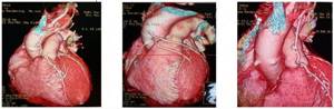
MSCT coronary angiography
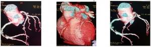
The heart is a moving organ. So that the vessels of the heart, the so-called. coronary arteries can be clearly distinguished; a special program for synchronizing the image with the heart rhythm is required. Synchronization with ECG ensures minimal heart motion and optimal image quality.
Along with assessing the condition of the coronary bed, multislice computed tomography of the coronary arteries (MSCT CA) allows one to study valve structures (cusp calcification, anomalies of valve development, vegetations), reveals myocardial lesions (scars, aneurysms, hypertrophies, ruptures), the condition of the cavities of the heart and pericardium. Additional information is provided by determining the systolic function of the myocardium with identifying areas of impaired contractility. Modern MSCT devices have the opportunity to study myocardial perfusion and its viability.
The “gold standard” for diagnosing coronary artery diseases is the so-called. invasive coronary angiography, in which, however, it is possible to evaluate only the lumen of the arteries. With atherosclerotic damage to blood vessels, one can see a narrowing or expansion of the lumen, but cannot in any way assess the structure and structure of the so-called. atherosclerotic plaques. For this purpose, an invasive method is used - intravascular ultrasound (IVUS). An important value of CA MSCT is the possibility of morphological assessment of the plaque, without the use of invasive intravascular examination. The accuracy and comparability of measurements of the degree of narrowing of the arteries during multislice computed tomography of the coronary arteries (MSCT CA) with IVUS data was noted. In addition, a special computer program makes it possible to accurately assess the functional significance of the narrowing of the coronary artery, which until recently was only possible using invasive methods.
Multislice computed tomography of the coronary arteries
- Cost: 16,000 rub.
More details
The 3D model obtained from image reconstruction is indispensable in identifying anomalies in the development of coronary arteries, anomalies in the development of the heart and other large vessels of the heart, and arteriovenous fistulas. MSCT of the coronary arteries provides important information for the interventional cardiologist in the presence of complete blockage of the lumen of the coronary arteries, the so-called. chronic occlusions, allowing to obtain additional data necessary to perform recanalization of the affected arteries. Thus, multislice computed tomography of the coronary arteries (MSCT CA) combines the capabilities of several diagnostic techniques: coronary angiography (CAG), echocardiography, cardiac MRI, and IVUS. Without exaggeration, we can say that, based on the totality of data, the MSCT CA technique for studying the heart and its vessels has no equal.
MSCT shuntography
After undergoing coronary artery bypass surgery, the condition of aorto- and mammary-coronary bypass grafts is of fundamental importance not only for assessing the quality of life, but also the prognosis of patients.
The MSCT method allows you to clearly visualize the number of shunts, their place of attachment and functional state with minimal X-ray exposure and a minimal amount of contrast agent administration.
MSCT shuntography is a highly accurate, effective and safe method for diagnosing pathological changes in the coronary bed in patients who have undergone coronary bypass surgery; this method is an alternative to traditional coronary angiography and shuntography, and taking into account the above, it is more preferable.
MSCT of brachiocephalic arteries
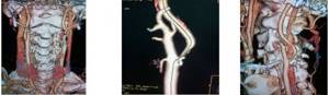
The brachiocephalic arteries are responsible for supplying blood to the arteries of the upper limbs and the brain.
CT angiography of the brachiocephalic arteries is a fairly new method in the diagnosis of vascular lesions in Russian medical practice. With a limited number of contraindications to the study, this method allows you to obtain an exceptionally clear picture of the vascular bed, both in two- and three-dimensional projection, and correlate it with bone structures.
It is impossible to imagine the work of a vascular surgeon without studying CT angiography data of the brachiocephalic arteries. Vascular interventions, whether open surgery or stenting, are impossible without a preliminary accurate diagnosis. CT angiography allows you to make all the necessary measurements and calculations to select the right stent and, in the shortest possible time, carry out stenting of a stenotic artery, plan an operation for pathological tortuosity of the artery, anomalies or atypical development of brachiocephalic vessels. In this case, as with MSCT coronary angiography, it is possible to evaluate atheroclerotic plaque or other causes of compression or narrowing of the vessels supplying the brain and upper limbs.
Indications and contraindications
MSCT is prescribed by a doctor in case of a history of renal colic, as well as persistent pain in the lumbar region. If pathological impurities are detected in the urine (protein, blood, uric acid crystals, increased number of leukocytes). If the cause of renal failure is clarified.
Contraindications to the service are intolerance to radiopaque (iodine-containing) drugs, severe renal failure. In children and pregnant women, the study is carried out according to strict indications, since the procedure is accompanied by ionizing radiation.
MSCT cardiography
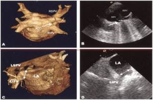
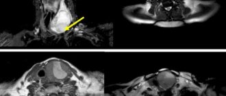
Accessory right pulmonary vein (arrow) identified by MSCT (A) and transthoracic echocardiography (B). Common ostium (double arrow) of the left pulmonary vein identified by MSCT (C) and transthoracic echocardiography (D)
In recent years, radiofrequency catheter ablation (RFA) techniques have been increasingly used to treat cardiac arrhythmias. With atrial fibrillation (atrial fibrillation), the focus of the chaotic rhythm is in the left atrium, at the mouth of the pulmonary veins. Knowledge of the anatomy of the left atrium is necessary before the planned intervention. MSCT cardiography provides a unique method for assessing the volume of the left atrium, as well as determining the individual anatomical structure of the pulmonary veins, determining the number and diameters of the mouths of the pulmonary veins and the angles at which the veins flow into the atrium.
This is an indispensable study when planning RFA surgery around the ostia of the pulmonary veins.
The operation is performed in patients with atrial fibrillation. Catheter ablation of the pulmonary vein ostia remains the only minimally invasive procedure with a proven effect on the anatomical substrate of arrhythmia.
Indications for MSCT of the kidneys
Any type of CT scan involves radiation exposure to the body. When performing MSCT, the radiation dose is minimal and not hazardous to health, however, this study is not performed without indications. Doctors usually give a referral for multislice tomography in the following cases:
- For symptoms that usually accompany disorders of the adrenal glands, kidneys and ureters (pain in the back and lower back, mucus and blood in the urine, swelling, painful urination, etc.).
- In case of lower back injury, when there is a possibility of kidney rupture.
- With obvious signs of hormonal imbalance.
- If the development of tumors and cystic formations, vascular lesions and inflammatory processes is suspected.
Typically, MSCT is an additional examination method. This procedure is recommended if questionable results of excretory urography or ultrasound are obtained.
Regular examination is recommended for patients with chronic kidney and adrenal diseases. It makes it possible to track the dynamics of the disease with high reliability and efficiency.
MSCT aortography
If the aortic valve is damaged, the only treatment method is its replacement. Traditionally, this has been performed using an open surgical approach. In recent years, endovascular prosthetics has been increasingly used in patients of the older age group. The principle of the operation of endovascular aortic valve replacement is that through an access through the femoral artery or a mini-access to the apex of the heart (also called apical), a special catheter with an artificial valve placed at its end is brought to the site of the damaged aortic valve. Before installing this artificial valve, the lumen of the own aortic valve is expanded and endovascular implantation of a valve selected to size is performed.
A distinctive feature of this treatment option is the low incidence of postoperative complications, minimal recovery time for the patient after surgery, and the possibility of performing it in patients with severe concomitant diseases. MSCT aortography before intervention is a prerequisite for aortic valve replacement. The method is used to plan traditional or endovascular aortic valve replacement surgery. MSCT aortography allows you to reliably measure the diameters of the aortic valve ring, calculate the diameter of the aorta, arteries necessary for surgical or endovascular access, as well as in the selection of the necessary instruments.
Dilatation and dissection of the aorta—aortic aneurysm—is a life-threatening condition and requires surgery. MSCT allows you to “see” the entire aorta with all its branches, the cavity of the aneurysm, its extent, its relationship with neighboring organs, and much more. In terms of information content for the surgeon, MSCT aortography or pan-ortography is a completely unique technique.
The MSCT method is indispensable for diagnosing aortic dissection and aneurysms and individual planning of minimally invasive surgery in the shortest possible time.

Aortic dissection is a complex problem in modern cardiovascular surgery. To date, for both acute and chronic aortic dissection, endovascular treatment has shown high effectiveness; for distal aortic dissection, it is the least traumatic and at the same time the most promising and is currently being actively developed. The principle of endovascular surgery is the implantation of an intravascular prosthesis (endoprosthesis), which allows isolating the dilated part of the aorta from the blood flow, preventing its rupture.
MSCT makes it possible to fully plan and calculate the size of the endoprosthesis individually for each patient.
Calculation of the size of the endoprosthesis for aneurysms of the thoracic and abdominal aorta is carried out according to a universal protocol based on MSCT data of the aorta.
In the postoperative period, MSCT of the aorta is the main method for assessing and monitoring the condition of the stent graft, in the early diagnosis of complications of endoprosthetics, and much more.
MSCT angiography of the arteries of the lower extremities
Patients with atherosclerosis and diabetes mellitus often experience severe damage to the arteries of the lower extremities. Intermittent claudication, trophic changes in the skin, ulcers, secondary infections, impaired sensitivity and motor function, up to gangrene, with the need for limb amputation - this is an incomplete list of the suffering of these patients. The only possible radical treatment for such patients is open surgery or endovascular surgery to restore blood supply.
MSCT angiography allows a detailed assessment of the condition of the vascular bed and provides invaluable assistance in planning surgical treatment. The study is carried out in case of damage to the vessels of the lower extremities to identify aneurysms; hemodynamically significant narrowing (stenosis) and occlusion of the iliac, femoral arteries, and arteries of the leg. After surgery, MSCT angiography is necessary to monitor the patency of vascular stents or shunts; condition of vascular prostheses, as well as, if necessary, planning re-interventions.
Features of the tomography procedure
After entering the X-ray room, the patient is placed on a movable tomograph couch. Then the laboratory assistant leaves the room and observes the procedure through a special window in the computer room. The tomography machine is equipped with an intercom, and in case of discomfort and deterioration of his condition, the patient can always report this. At the doctor's command, the patient will need to hold his breath for a few seconds during the scan.
The examination is carried out painlessly, quickly and comfortably. Native scanning takes 3-5 minutes. Contrast increases the duration of screening by 20 minutes. Results are provided in most medical centers in St. Petersburg on the same day within 30-60 minutes. The conclusion obtained in the process of describing the results of a CT scan of the adrenal glands is taken into account by the attending physician to establish a diagnosis and subsequent treatment.
Leading doctors of St. Petersburg

Pashkova Anna Alexandrovna
specialization: MRI doctor, radiologist of the highest category, candidate of medical sciences. Appointment - at the AMI Medical Center, Standard MRI on Ladozhskaya
medical experience - 21 years
Online registration
Khalikov Aziz Jalanovich
specialization: MRI doctor, radiologist of the highest category, candidate of medical sciences. Registration - at MC Scandinavia
medical experience - 20 years
Online registration
Trufanov Gennady Evgenievich
specialization MRI doctor, MD, professor. Recording - Federal State Budgetary Institution "National Medical Research Center named after. V. A. Almazova"
medical experience - 35 years
Online registration
Kholin Alexander Vasilievich
specialization MRI Doctor Professor of the Department of Radiation Diagnostics Northwestern State Medical University Appointment - CMRT Staraya Derevnya, CMRT Petrogradskaya
medical experience - 35 years
Online registration