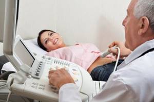
Benefits of Ultrasound
How is ultrasound different from x-ray? These are two completely different methods of diagnosing patients.
Each of them has its own advantages and disadvantages. There are certainly more advantages with ultrasound. So how are they different? The most important difference is that ultrasound diagnostics is not an invasive method. This means that during the scan the body is not exposed to radiation. This is the main advantage of ultrasound. Over the years of improving this method, it can be used to examine even bone and ligament tissue.
The idea of using ultrasound is not new; the first scanner was invented back in the 70s. Later, this technology appeared with us, then our doctors were able to significantly increase their work productivity. In order to correctly make a diagnosis, it is necessary, firstly, to study the symptoms, and secondly, if necessary, special procedures are carried out, ultrasound is one of them. It is extremely useful, as it allows you to identify various ailments at an early stage of development.
This includes various oncological formations. The principle of operation is quite simple. A gel is applied to the area to be examined, then the doctor uses a special sensor to conduct the examination. The signal from the sensor is transmitted to the tissues of the desired organ, during which time the ultrasound penetrates various liquids.
Having reached its target, it is reflected back to the sensor, thus being displayed on the monitor screen.
Many people ask the question: what is better: X-ray or ultrasound?
To understand correctly, you need to know all the advantages and disadvantages of each method.
The feasibility of each method depends on the complexity of the clinical case, anatomical differences, structures involved in the pathological process, the need to confirm the final diagnosis, determine the stage of the disease, assess the effectiveness of the treatment and the safety of the chosen diagnostic method for the body as a whole. The best doctors in Kerch in the medical field will be able to competently advise you, prescribe and interpret tests, correctly refer you to the necessary additional diagnostic method, accurately diagnose and give effective prescriptions and recommendations for treatment.
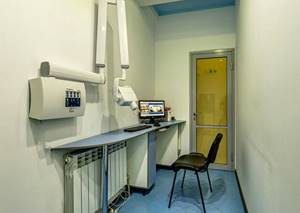
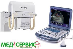
The largest private clinic in Kerch has a lot of modern medical diagnostic equipment of Expert and Premium class, which is absolutely available to you without queues or hassle.
Thus, often to check the condition of bone structures, teeth, joints, spine or respiratory organs, the latest digital X-ray machine Philips (Netherlands) or radiovisiographic system Soredex (Finland) is used, and to assess internal organs, the cardiovascular system, thyroid gland or reproductive system systems choose an innovative ultrasound scanner from GE Healthcare (USA) with 3D and 4D visualization capabilities and Doppler for assessing blood flow. To assess the functioning of the brain, in addition to those described above, the echoencephalometry method is used. A cardiologist often prioritizes ECG interpretation or 24-hour Holter monitoring of ECG and blood pressure. Gynecologists cannot do without a microscope and video colposcope in high resolution FullHD.
When planning operations to eliminate flat feet or hallux valgus, photographs are always taken in different projections, with and without a load. The dental department always uses radiovisiography of teeth, apex location to determine the topography of the root canal, orthopantomography, electroodonodiagnostics to clarify the vitality of the dental pulp, and much more. Thus, to clarify the diagnosis as quickly and correctly as possible, it is advisable to use a comprehensive diagnosis of organs and systems.

Despite the most powerful material and technical base of medical equipment that meets all requirements and standards, the most important place in the correct diagnosis is occupied by the doctor. Invaluable many years of experience and clinical thinking even when carrying out basic methods of examination and research, such as palpation (palpation), percussion (tapping), auscultation (listening) and the correct collection of complaints and anamnesis, the correct interpretation of tests, allows us to accurately assume the presence of a particular problem , which is only confirmed in consultation with related specialists and the implementation of additional methods. Remember, early or timely consultation with a doctor significantly increases the chances of correct diagnosis and effective treatment.
Diagnostics in Astana
It’s good that our citizens take care of their own health, improve medical literacy, lead a healthy lifestyle, etc. If there is a need for quality medical care, then contact us, Medar Clinic.
We have all the necessary equipment for high-quality diagnostics and treatment. More than ten years of experience, unique techniques, cozy rooms, friendly medical staff. We also help with rehabilitation after severe injuries or operations. Our patients are always satisfied with the results.
In our methods, we use psychological support for the patient, special diets, traditional medicine, physical therapy and the most advanced healing methods.

Ultrasound or X-ray
When any health problem arises, every person is faced with the question of which doctor to contact and which diagnostic method to choose.
It is, of course, worth starting with the fact that this should be decided by the attending physician, since differential diagnostic methods are used to identify different pathologies.
Among the most popular diagnostic methods today are ultrasound and x-ray diagnostics.
Vinikli nutrition - consult a doctor
To make an appointment for a consultation, call or fill out the return form:
(050) 301-99-26 (067) 446-11-79
Damn,
Your request has been successfully sent!
A call center specialist will contact you as soon as possible to clarify all the details.
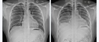
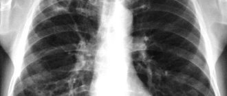
What does an x-ray show?
An oval lump can be seen in the image. This is the heart. It is located on the left side of the chest and is placed in a vertical direction (in people with excess body weight, the heart is slightly turned, which is associated with an increased load on it). Large main vessels are located just above this seal. The image can also see clearly defined arcs that represent the cardiac chambers, and if they are straightened, the patient is diagnosed by myocardial pathologists.
In addition, radiography allows you to see the following deviations:
- calcification of blood vessels and heart valves;
- anomalies in the development of the organ and its structures;
- changes in the structure of the aorta and pulmonary artery;
- damage to the tissues of the internal organs of the chest;
- violation of the location of the diaphragm;
- increase in the volume of the pericardial sac.
Difference between methods
The most important difference in these two methods of examination is the principle of operation of the active elements that provide a picture of the area under study.
Thus, during an ultrasound examination, an image is obtained by sending ultra-frequency waves or signals, some of which pass through the organ, and some return back, which makes it possible to display the image on the device’s monitor. The difference between the transmitted and reflected waves is called the reflection coefficient. It directly depends on the density and structure of the organ being studied. The coefficient is captured by a special sensor, which converts the waves into a picture on the monitor.
There are different types of resulting images - two-dimensional, three-dimensional and four-dimensional, which show a regular, volumetric and time-lapse image, respectively.
The principle of operation of x-rays is completely different. It consists in the impact of radiation waves on the part being studied. Penetrating radiation - X-rays - allows you to obtain images of organs and structural components of the skeleton. This method is called radiography. It is also possible to obtain images of organs on the screen in real time. This method is called fluoroscopy.
It is noteworthy that the soft tissues of the human body completely transmit the sent rays, while bones or solid foreign bodies, which for some reason end up in the body, delay these rays.
Since the radiation that is the basis of this diagnostic technique is ionizing radiation, this diagnostic method has some limitations (for example, the number of procedures is reduced to one per year or a ban on the use of X-ray diagnostics for pregnant women).
Recommendations
Pleural cavity:
▣ Pneumothorax
- Sonographic signs of pneumothorax: absence of lung sliding, absence of vertical reverberation artifacts, absence of pulmonary pulse sign and the presence of a pulmonary point (the point of contact of the visceral and parietal pleura in pneumothorax).
- The presence of pulmonary sliding artifacts and/or vertical reverberation emanating from the pleural line and/or pulmonary pulses excludes pneumothorax.
- In a patient with acute respiratory failure and a strong suspicion of pneumothorax, there is no need to look for a lung point.
- Lung ultrasound is a better diagnostic imaging modality than chest x-ray for patients with pneumothorax; however, pulmonary ultrasound is less useful than chest x-ray for making therapeutic decisions such as chest drainage.
- Convex and linear probes are recommended for diagnosing pneumothorax.
- The absence of a pulmonary point with the simultaneous presence of pneumothorax occurs in critical or closed pneumothorax.
- Previous pleurodesis influences the presence of lung sliding sign (the sign will be absent or limited) and vertical reverberation artifacts (artifacts arise from pleural line abnormalities). The presence of vertical artifacts excludes pneumothorax in patients undergoing pleurodesis.
- Localized pneumothorax – a pocket of pleural air can be visualized; the air in the pleural cavity does not move when the patient's position changes.
- The lung point is the boundary between the pleural air pocket and the normal pleural cavity; this sign can be visualized in B or M mode.
- The pulmonary pulse is the pulse of the lung resulting from cardiac motion transmitted to the lung; Pulmonary pulses are best visualized with M-mode and/or power/color Doppler parameters.
- The recommended position for examination is the supine position (except for patients with orthopnea).
▣ Pleural effusion
- Chest ultrasound is more sensitive and more specific for diagnosing pleural effusion than chest x-ray.
- The sensitivity of chest ultrasound in determining pleural fluid volume is similar to that of chest computed tomography.
- Ultrasound is a good method for visualizing the chest, which allows you to find the optimal place for puncture.
- Chest ultrasound helps minimize post-puncture complications.
- Sonomorphology of pleural fluid in combination with clinical data suggests its type.
- If possible, pulmonary ultrasound should be performed in any patient with a clinical suspicion of pleural effusion and/or when the classic radiological finding suggests the presence of pleural effusion, especially when thoracentesis is required.
- Thickening of the parietal pleura (more than 2 mm) and/or detection of focal lesions within the parietal pleura may indicate a metastatic type.
- Ultrasound assessment of the volume of free fluid in the pleural cavity is possible using mathematical formulas. Below are some examples of formulas depending on body position:
- Sitting position: V (ml) = LH (cm) × 90 or V (ml) = [LH (cm) + SH (cm)] × 70; measurement along the posterior axillary line, LH – height of the fluid layer, SH – average distance between the diaphragm and the base of the lung
- Supine position: V (ml) = T (mm) × 20; exhalation measurement; T – thickness of the liquid layer.