Story
Chinese doctors were the first to describe in detail the procedure for facial reconstruction for cleft lip disease.
Early techniques involved cutting a straight slit and sewing it together piece by piece. In the mid-17th century, the evolution of surgical techniques allowed the use of special plates to treat such diseases. The use of this method in the early stages of a child’s life is the basis of reconstructive interventions even now. Tennyson introduced the method of triangular skin flaps, which forms a Cupid's bow as a result of surgical intervention on the upper lip. The geometry of this technique was described in the works of Randall, who considered it the most popular at that time. Millard introduced a surgical technique that involved moving the medial portion of the lip toward the lateral plate while maintaining the cupid's bow shape of a deep groove. His theory contributed to improving the results of plastic surgery for cleft lip .
1.What is reconstructive microsurgery?
Reconstructive microsurgery is a branch of surgery that deals with the restoration of deeply damaged tissues. Reconstructive microsurgery can be used both for the treatment of tumors or organ transplants, and for the purposes of aesthetic medicine.
Reconstructive microsurgery is often used to treat congenital pathologies and deformations of the structure of the human face and body. One of these pathologies is the so-called. "cleft lip" or "cleft palate".
A must read! Help with treatment and hospitalization!
Issues
Cleft lip is one of the most common congenital pathologies. This condition is caused by insufficient development of mesenchymal cells during the primary formation of the palate. This occurs from the fourth to the seventh week of fetal development. The consequence of this development is distortion of the upper lip and nose. Also, cleft lip may be one of several related syndromes, such as facial development deficiency, dental anomalies, and speech impairment (in the presence of a cleft palate). Such children very often have communication difficulties in later life associated with their psychological state.
Cleft lip and palate: the defect can be corrected
Cleft lip and palate is one of the most common congenital malformations of the upper jaw: it occurs in a third of patients with congenital malformations. In Moscow, 1 in 800 babies are born with this developmental defect.
The anomaly is usually detected by ultrasound in the 1st-2nd trimester of pregnancy. Such news is a huge stress for future parents. But in most cases, thanks to timely and qualified treatment, cosmetic and functional defects in the baby can be eliminated.
At what age is surgical treatment most effective and how a complex anomaly is treated, Elvira Sergeevna Mkrtumyan, a maxillofacial surgeon and plastic surgeon at the Morozov Children's Hospital, told.
What influences the formation of a defect?
The defect is formed in the first two months of the child’s intrauterine development, when the formation of the facial skeleton occurs. The cause of the development of pathology is a chromosome abnormality.
The development of the defect is influenced by both hereditary factors (7%) and external factors (40%). External ones include infectious and viral diseases, smoking, drugs, alcoholism, the use of certain medications, lack of vitamin folic acid, poor ecology, and so on.
What types of clefts are there?
Clefts can occur in both the lips and palate. They, in turn, can be isolated (only the lips or only the palate) and through, when the defect extends to the upper lip, alviolar process and palate. Each type has varying degrees of severity. In addition, the defect can be either unilateral (usually on the left) or bilateral.
With an isolated cleft lip, as a rule, the deformation may be minor, or may be accompanied by deformation of the nasal septum, alar cartilage and alveolar cleft, which subsequently leads to dental problems.
With cleft palates (from minor clefts of the soft palate only to severe bilateral lesions of the soft and hard palates), children may have problems with eating, breathing, sucking, and subsequently with sound pronunciation. Thanks to timely surgical intervention, it is possible to obtain an ideal treatment result.
The most severe form is bilateral through cleft lip and palate. But even in this case, timely treatment and rehabilitation give the baby a chance for recovery.
If, during intrauterine diagnostics, a cleft is discovered in the baby, what should parents do?
The most important thing I would like to say to parents who are faced with this problem is that there is no need to worry. It is important to immediately consult with maxillofacial surgeons involved in the treatment of pathology.
We advise future parents even before the baby is born. We tell you how to feed your child correctly - it is better with a spoon or a bottle with special nipples in order to preserve swallowing reflexes, because with prolonged tube feeding they can fade.
Often this defect can be accompanied by pathology of the bone and cardiovascular systems, kidneys. Therefore, it is important to undergo tests immediately after birth to rule out these diseases. If the baby does not have other serious concomitant diseases, we prescribe planned surgical treatment to eliminate the defect.
At the request of the parents, after the maternity hospital, the child can be transferred to the neonatology department of our hospital, where the mother will be taught how to feed the child, examined and given recommendations for management before a planned operation.
At what age is surgical treatment most effective?
The optimal age for surgery to correct a cleft lip and eliminate nasal deformity, if there is no gross pathology among other systems, is 3 months. Earlier plastic surgeries do not provide the opportunity to obtain the best aesthetic result.
If the child has a severe form of cleft, correction can be carried out in two stages. At six months, if it concerns the palate, it is the soft palate; from one to two years, it is the hard palate. If surgical treatment is carried out in one stage, then it is performed at the age of 9 months to 1.5 years. This has a number of advantages: the child endures one rather than two anesthesia, the tissues are more mature, which means the treatment result will be more effective. We recommend palate plastic surgery for children under two years of age, until the child’s brain—the center of speech—has formed incorrect speech.
If the child has a severe form of cleft, correction can be carried out in two stages. At six months, if it concerns the palate, it is the soft palate; from one to two years, it is the hard palate. If surgical treatment is carried out in one stage, then it is performed at the age of 9 months to 1.5 years. This has a number of advantages: the child endures one rather than two anesthesia, the tissues are more mature, which means the treatment result will be more effective. We recommend palate plastic surgery for children under two years of age, until the child’s brain—the center of speech—has formed incorrect speech.
What operations are performed for cleft lip and palate?
Correction of a congenital anomaly requires an integrated approach. It is aimed not only at eliminating a cosmetic defect, but also at reconstructing functional disorders, eliminating problems with swallowing, breathing and sound pronunciation. The volume and type of operational benefit depends on the characteristics of each child and is selected individually.
If the cleft looks like an isolated lip or palate, surgical treatment will be performed in one stage. In the most difficult cases, correction is carried out in several stages.
For isolated cleft lip without significant deformation of the nose, cheiloplasty is performed. The vestibule of the oral cavity (the space located under the upper lip) is created, the integrity of the orbicularis muscle of the upper lip is restored, and a red border is formed. If the upper lip defect is more severe, then a one-stage cheilorhinoplasty (lip and nose surgery) is performed. We perform lip plastic surgery and place the alar cartilages of the nose in a symmetrical position. On the 7th day, postoperative sutures are removed and the children are discharged from the hospital with recommendations for further management of the child.
An isolated cleft palate manifests itself as an anatomical damage to the soft palate (mobile palate) or the soft and hard palate. The pathology is accompanied by impaired swallowing, breathing and sound pronunciation. Palate plastic surgery involves restoration of the hard palate and soft tissues of the velum, and closure of the alveolar process. After the operation, when children reach 3 years of age, classes with a speech therapist are recommended to prevent speech disorders, manifested by nasality and unintelligibility of speech.
If speech disorders are not eliminated through the efforts of speech therapists alone, then there is a surgical correction - elimination of velopharyngeal insufficiency. The operation involves transplanting flaps on a pedicle in the area of the pharyngeal ring.
From the age of 5, a child is sent for treatment to an orthodontist to eliminate defects in the dentofacial system. At the end of orthodontic treatment, the next stage of surgical treatment is performed - bone grafting of the alveolar cleft to eliminate the bone defect of the alveolar process.
The Morozov Children's Hospital has accumulated extensive experience in the effective treatment of cleft lip and palate. Every year, doctors perform up to 200 operations to correct these congenital malformations.
Spreading
Among the fair-skinned population, only one child is born with this pathology out of 1000 newborns. Cleft lip is twice as common in Asia. The Negroid race is characterized by the presence of this disease in half of the population. Most often, cleft lip occurs in boys. Isolated clefts on the right side can be found half as often as on the left side, and those in turn can be found 9 times more often than with bilateral cracks. Of the total number of children suffering from this disease, 50% are carriers of combined cleft (lip and palate), 30% - isolated (palate only), and 20% have alveoli and cleft lips. Less than 10% of the settlements are bilateral cracks.
The risk of congenital pathology in the next generation, if one of the parents has a combined cleft, is 4%. If two parents have a cleft lip, the chance of inheriting the disease increases by 9%.
What to do if an ultrasound reveals abnormalities of the fetus’s face or jaws
Suspicion of a serious pathology always puts parents in a state of shock. They have to decide whether to continue the pregnancy.
If your child has a cleft lip, you should not sign up for an abortion. In the first year of life, the baby will be operated on and he will not be different from other children. In other cases requiring multi-stage plastic surgery, you need to consult with a surgeon about the success of such interventions. If a woman nevertheless decides to have an abortion, it is better to terminate the pregnancy at a short term using medication.
Since such pathologies can be either independent or a symptom of severe hereditary diseases, the woman is prescribed additional examinations - analysis of amniotic fluid, blood from the umbilical cord, and placental tissue.
If the examination shows that the child will be born with a serious hereditary illness, parents need to decide whether to give birth to a sick baby. Everyone answers this question for themselves, but in any case it wouldn’t hurt to listen to the opinion of doctors.
If you find an error, please select a piece of text and press Ctrl+Enter
Etiology
The fact that the appearance of an isolated cleft in a child is influenced by teratogenic agents has not been sufficiently studied. An exception is the anticonvulsant drug phenytoin. Its use during pregnancy is associated with a tenfold increase in the incidence of babies born with cleft lip. A child with a characteristic pathology is born to a non-smoking mother two times less often than to a smoking woman. The presence of dislocation is associated with disturbances in the functioning of other organs in 5-14%.
Vanderwood syndrome is the most common cleft palate and lip disorder. This autosomal dominant disorder is characterized by a cleft lip (palate) and paranasal sinus, or pit in the lower lip. A cleft palate is more often associated with the syndrome than an isolated fissure of the lip or a combined cleft. In most cases, cleft lip only is considered to be the result of many factors or a change in a gene in the chromosome, but not a sign of Vanderwood.
FREE CONSULTATIONS!
We are pleased to announce the start of free admission at the Konstanta Clinic for patients under 18 years of age by maxillofacial surgeon, pediatric surgeon L.A. Eremeyshvili. for questions:
- Congenital facial pathologies: cleft lip and palate;
- Benign neoplasms of the face and oral cavity;
- Diseases of the temporomandibular joints;
- Pathology of the frenulum of the oral cavity: lips and tongue.
Correction (plasty) of tongue frenulum is provided free of charge for children under 1 year of age.
*Valid for residents of Yaroslavl and the Yaroslavl region.
You can make an appointment by phone or through the website (4852) 37-00-85 Daily from 8:00 to 20:00
Sign up for a consultation
Congenital cleft lip or “cleft lip” is one of the most common developmental defects, statistically ranking 3rd among all genetic abnormalities. The detection rate is at the level of 1:700 newborns.
Classification
There is no generally accepted classification of cleft lip and palate. However, 4 groups are clearly defined:
- Clefts of the soft palate only
- Clefts of the soft and hard palate
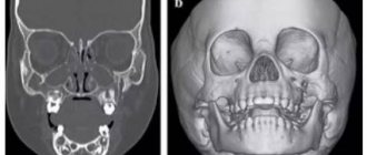
- Complete unilateral cleft lip and palate
- Complete bilateral cleft lip and palate
This group does not apply to isolated cleft lips and incomplete fissures. The Kernahan classification differentiates cleft lip, palate, and alveoli. The boundary between the clefts of the primary and secondary palate is determined by sharply defined openings.
Performance
Cleft lips appear in the form of microforms (complete and incomplete cracks). They are characterized by a vertical groove and a scarlet notch with varying degrees of lip reduction. An incomplete unilateral cleft lip of varying degrees is manifested by disruption of the base of the nose, covering the range from the edge of the lip to the nostril. A complete cleft lip, in addition to the signs already listed, is characterized by an alveolus.
Bilateral clefts are almost always associated with the palate. 86% of patients with this pathology have a fissure of the palate. Unilateral cleft lips are associated with palatal abnormalities in 68% of cases. Pushing food through the nasal cavity while eating indicates the presence of a cleft palate. Children with a cleft lip must undergo a complete examination of the head, neck and palate to the very tip of the tongue. The presence of a bifurcated uvula, which is a translucent palatal curtain in the central part and a noticeable gap on the posterior border of the hard palate, indicates a submucosal palatal cleft.
All newborns with cleft lip suffer from chronic otitis media or effusion of the middle ear, which is inextricably linked with the palatal cleft and requires constant care. To solve this problem, otolaryngologists resort to a myringotomy procedure, which can be performed before or immediately after treatment for Vanderwood syndrome.
In most cases, a cleft lip is not a death sentence. Parents of such children must understand that their situation is not critical. When you first visit your doctor, they can find out detailed information about feeding methods for cleft lip. For a child with this pathology, the ability to suck milk is difficult. However, with an isolated cleft lip and alveoli, breastfeeding is quite possible. Special bottles and nipples have been created to feed children with a cleft palate. Transverse soft nipples help premature babies with cleft palate.
Advantages and disadvantages of 2, 3, 4D fetal ultrasound
Two-dimensional ultrasound during pregnancy has significantly better diagnostic capabilities. Despite the fact that the images of such an ultrasound look unpresentable, it, unlike three-dimensional, allows the doctor to judge not only the external data of the person, but also the degree of development of his internal organs and systems.
It is important to know that 3D/4D ultrasound is performed in addition to regular fetal ultrasound and cannot replace it. Only for some malformations does the use of “volumetric” ultrasound provide the doctor with additional information.
What advantage does 3D have over 2D ultrasound images? From a medical point of view, none at all. Only in some cases is 3D ultrasound indicated. The main and decisive difference between a classic black and white ultrasound image and three- and four-dimensional images is that some specialists can see in three-dimensional images what an expert sees in two-dimensional ones.
There are limitations for 3D ultrasound due to gestational age and the position of the fetus in the womb. If there are indications for 3D ultrasound, then the optimal period is considered to be 20-24 weeks of pregnancy. During this period, all external organs of the fetus are formed, and the doctor has the opportunity to identify pathology of the facial skeleton (“cleft lip”, “cleft palate”). But if the baby turns his back to the sensor, his face will no longer be visible.
Although ultrasound imaging has been around for many years, pregnant women and their families need to know that the long-term and repeated effects of ultrasound on the fetus are not yet fully understood.
Thus, the FDA, the American Federal Agency for Control of Food and Drugs, strongly opposes the use of three-dimensional or four-dimensional ultrasound (3D ultrasound, 4D ultrasound) for commercial purposes, since prenatal ultrasound examinations cannot be considered absolutely harmless. Trying to capture the best frame or video, the FDA notes, doctors increase the power of the emitter, forcing pregnant women to spend an unacceptably long time under the influence of the device - 30 minutes or more, which can affect the health of the unborn newborn.
Ultrasound is a form of sound waves that vibrate approximately one hundred times faster than normal sound waves. Ultrasound waves can damage human tissue in a variety of ways. For example, high temperature is one of the effects. In addition, although ultrasonic waves do not produce audible noise, secondary vibrations do produce noises that can cause fetal movement. Other damaging effects include the formation of tiny bubbles in tissue, changes in interstitial pressure, indication of flow within fluids, and the formation of small amounts of toxic substances.
Functional anatomy
A complete unilateral cleft lip results from a typical deformation of the soft tissue underlying bony and cartilaginous structures. Muscular imbalance affects the maxilla by rotating the mesial incisal and posterolateral portions.
The lower edge of the anterior nasal septum is displaced from the vomerine cleft into the intact nostril and rests on the cleft from the sides. The columella, which lies above, does not reach the sides of the septum and is distorted due to the movement of the caudal septum. At the tip of the nose, the axillary cartilage is deformed, and the medial crus moves backward. The fornix is separated from the uninjured side, and the lateral crus is flattened and extended through the gap. The nasal axis from the side of the cleft is located in the horizontal plane. On the opposite side, the nostril axis is different and is located in the normal vertical plane.
The rounded muscle tissue is not located transversely, as in normal lips, but goes upward at an angle parallel to the edge of the cleft of the axillary base on the lateral side and is distributed to the base of the medial columella. The septum on the side of the cleft is very short and the supposed tip of Cupid's bow is higher. The presence of a bright red tint indicates an incomplete cleft on the medial side.
Complete bilateral cleft lip occurs as a result of failure of the incisal part and fusion with the lateral segment of the jaw. Subsequent growth of the incisal bone raises the vomer even higher, moving the protrusion beyond the side. In an isolated lip border with vertically shortened skin, the white roll is not sufficiently developed and the cinnabar is not bright enough. The central protruding part of the upper lip lacks muscle tissue, as well as the philtrum and dimple. Cupid's bow is missing. Bilateral nasal cleft deformity is characterized by a distinct pterygoid base and a wide separation of the dome portion of the axillary cartilage. The columella is significantly shortened, so it causes the nasal tip to droop.
How defects of the face and palate of the embryo are formed
A two-week-old embryo already has a primary oral fossa at the site of the future mouth, which gradually deepens. By the end of the first month, this area is limited to five tubercles - the frontal, two maxillary and two mandibular. From them the face and jaw apparatus are subsequently formed. This process ends around the seventh week. The palate is formed by 10-11 weeks.
Adverse effects on the fetus before 11 weeks lead to the formation of birth defects.
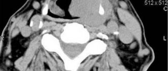
Anomalies in the development of the maxillofacial area are visible already at the first ultrasound screening, carried out at 11-14 weeks. However, some pathologies are detected during ultrasound examinations at a later date.