Arteriography of the lower extremities is a method of visualizing the lumen of the arteries by introducing a radiopaque substance into them through a catheter and simultaneous fluoroscopy (video recording of x-rays), with recording and processing of the resulting image using special equipment. In this case, the specialist receives objective information about the anatomical structure of the vascular bed of the area under study, the speed of blood flow, the presence of stenoses (narrowings) and occlusions (complete blockage), the degree of development of collateral circulation in the limb under study.
Based on this information, a council of doctors sets indications for surgery and determines the stages of treatment; taking into account the entire complex of preoperative examination, the choice of revascularization method is made (open bypass, endovascular or hybrid intervention).
Treatment under the compulsory medical insurance policy is free!
Submit your application
Follow the news, subscribe to our social networks
Details
Advantages of angiography at the Innovative Vascular Center
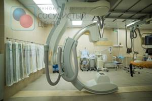
Arteriography of the lower extremities is performed on a stationary Philips Allura Xper FD20 angiographic complex, or on a Veradius mobile angiograph if performed intraoperatively. The use of modern angiographic systems makes it possible to conduct research at any required frame rate, which gives an objective picture of the nature of blood circulation in the limb.
Our clinic uses CO2 angiography without the use of iodine contrast. This method is well suited for patients with iodine intolerance and allows any operation to be performed using the endovascular method.
To reduce the risk of complications, we perform angiography most often through a peripheral radial approach on the arm, which allows the patient to begin walking immediately after the procedure.
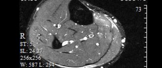
What is diagnosed using a tomograph?
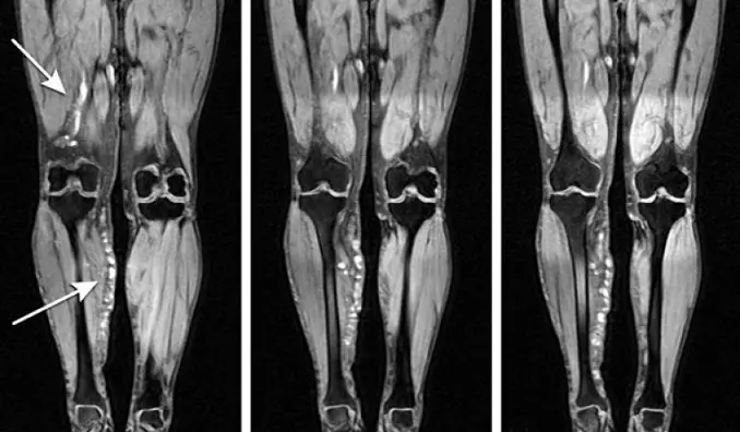
Unlike X-ray studies, which clearly visualize bone structures, MRI is more aimed at studying soft tissues. Based on the detection of certain chemical elements, magnetic resonance imaging best visualizes areas saturated with water, and more precisely, hydrogen.
The photographs clearly show:
- muscles;
- fat deposits;
- large vessels and nerves;
- The lymph nodes;
- joint cavities.
In addition to healthy organs and tissues, pathologically altered areas, areas with inflammatory processes, and tumor growths in the lower extremities are easily detected.
In particular, the following diseases and pathological changes are diagnosed:
- congenital developmental anomalies;
- injuries and consequences of traumatization (scars, adhesions);
- degeneration of nerve fibers;
- violations of the shape of joint surfaces;
- enlarged lymph nodes;
- primary tumor sites and metastases;
- local inflammation.
With the help of a magnetic field, the signs of the listed pathologies are revealed much more clearly than when a computed tomography is performed using an X-ray machine. In addition, MRI is virtually harmless to the patient, while CT is not recommended to be repeated after a short period of time.
How is angiography of the lower extremities performed?
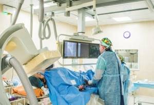
Angiography is performed by a highly qualified x-ray endovascular surgeon in a sterile cath lab and with sterile consumables. Anesthesiological monitoring is carried out throughout the study.
Under local anesthesia, a puncture of the artery of the arm or leg is performed, after which a thin catheter is inserted into its lumen, through which a radiopaque substance is injected. During the examination, the patient may feel warmth as the contrast is injected, and very rarely there may be pain. The duration of the angiography itself is 10-15 minutes.
When performing angiography, the specialist receives objective and accurate information about the structure and damage to the arterial bed of the area under study. The resulting images are saved electronically and can subsequently be written to disk and given to the patient.
Magnetic resonance therapy
Magnetic resonance imaging is a diagnostic procedure that allows you to obtain images of any part of the human body in any plane with the highest contrast of soft tissues.
This is a highly informative and at the same time safe method for studying human organs and systems, based on recording a radio signal received from hydrogen protons placed in a constant magnetic field.
About the tomograph:
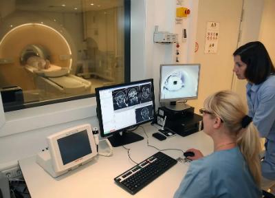
Our Center has a TOSHIBAVantageTitan 1.5 Tesla , a new generation high-tech medical device with a wide tunnel aperture, which makes the examination comfortable for the patient. The wide aperture allows magnetic resonance imaging to be performed on large patients and patients with claustrophobia.
The tomograph has a wide and short tunnel, it has lighting and a button for emergency communication with a doctor. During the study, medical personnel monitor the patient in the device.
The principle of image acquisition, unlike CT, is not associated with ionizing radiation; therefore, patients do not face radiation exposure. However, the strong magnetic field created by the tomograph imposes some limitations.
Absolute contraindications to MRI:
- Pacemakers, insulin pumps and other devices that affect the body (rhythm regulation, drug administration, etc.),
- First trimester of pregnancy,
- The presence of metal clips installed during surgery and other metal foreign bodies on the vessels of the brain or vessels of the eyes and orbits,
- The period is less than 6 months after the installation of shunts and stents in vessels, vena cava filters (containing metal).
The following studies are performed in the MRI room:
MRI of the head:
- brain,
- pituitary,
- orbits,
- paranasal sinuses,
- vessels of the brain and vessels of the neck.
MRI of the spine:
- cervical region,
- thoracic region,
- lumbosacral region,
- coccyx.
MRI of joints: knee, shoulder, hip, ankle, feet.
MRI of the pelvic organs (male and female);
MRI of soft tissues of the neck;
MRI of soft tissues of the lower extremities;
MRI of the abdominal cavity and retroperitoneal space.
All doctors working in the department have the highest certification category.
Complex diagnostic cases are brought up for discussion within interdisciplinary councils with the participation of doctors from different specialties.
All our doctors use the Archimedes medical information system, in the archive of which the data of all studies is stored for a long time, which allows us to assess the patient’s condition over time during repeated visits.
The duration of the study takes from 20 to 60 minutes, depending on the area of study and the complexity of the detected changes.
Some types of MRI examinations may require additional preparation. We talk about this separately when registering for the study.
Difference between CT and MRI:
CT and MRI are two types of studies that provide the most complete picture of the condition of organs and systems. Each method has its undeniable advantages.
MRI is safe and informative in diagnosing diseases of the central nervous system of the spinal cord, vascular bed, soft tissues, and joints.
CT is accurate and gives a detailed picture of injuries, for example, traumatic brain injury with suspected hemorrhage, diseases of internal organs (lungs, digestive system, genitourinary system and others), bleeding. However, CT does not have such a high degree of safety. The question of prescribing one or another type of examination is decided by the doctor, depending on the results of the preliminary diagnosis.
Appointments in the MRI and MSCT rooms are by appointment only.
Phone numbers for recording; +7 (499) 553-69-79
Opening hours: Monday-Friday from 9:00 to 19:00
Possible complications of angiography
Complications after angiography are very rare. However, due to the fact that this study is invasive (penetrating through the natural external barriers of the body) and is accompanied by the introduction of a radiocontrast agent into the systemic bloodstream, associated complications may still develop: contrast-induced nephropathy,
- bleeding
- hematomas and false aneurysms at arterial access sites
- local infectious complications
- allergic reactions to contrast
Our specialists do everything possible to prevent these complications, and also know the necessary treatment methods in case of development.
How is an MRI of the head and neck performed?
MRI of the head in Moscow is performed using modern equipment - a magnetic tomograph. 20-30 minutes is the average time it takes for a head MRI. The price of the study varies depending on the area of the skull being examined, as well as the status of the medical institution and the equipment used.
Once a person has decided where to get an MRI of the head, he does not have to worry about further preparation:
1) At a consultation with a specialist, the doctor gets to know the patient, his diseases, and allergic reactions.
2) The next step is to remove and remove from your pockets all metal and magnetized objects (watches, belts, bank cards, jewelry, items of clothing, keys, coins, glasses) during scanning. When performing an MRI of the head in Moscow, many clinics provide the patient with a change of clothes for the convenience of the procedure.
3) Having prepared, the person lies down on the table, which will move inside the tomograph body. To obtain the highest quality layer-by-layer image of the structures of the skull, it is necessary to lie as still as possible, taking a comfortable position in advance. After scanning, the table slides back out of the machine body.
Before you find out how much an MRI of the head costs and plan the procedure, you need to know an absolute contraindication: magnetic examination is not performed for people who have metal and magnetic objects implanted into their bodies (pacemakers, vascular implants, metal clips on brain vessels).
In all other controversial cases, the decision to conduct an examination in this way is made individually:
- Pregnancy (first trimester).
- Claustrophobia, nervous disorders.
- Severe health conditions.
- The presence of prostheses and implants that are not the reason for an absolute ban on the procedure of magnetic examination of the skull.
Price for ultrasound of blood vessels of the lower extremities (veins and arteries of the legs)
In the price list of services for ultrasound examination of veins and arteries of the lower extremities (vessels of the legs), you can compare all prices for ultrasound and select the necessary type of examination for you. If you need more detailed advice on the cost of ultrasound diagnostics or recommendations for examination, you can call us at 8 or make an appointment online on the website.
| Service | price, rub. | Promotion price, rub. | Record |
| Triplex/duplex scanning of vessels of the lower extremities (veins of the legs) with functional tests | 2100 rub. | Sign up | |
| Triplex/duplex scanning of the arteries of the lower extremities (arteries of the legs) with functional tests | 2100 rub. | Sign up |
Ultrasound diagnostics are performed by experienced doctors
Grebennikov Sergey Aleksandrovich, 25 years of experience
Position held in the clinic: Ultrasound doctor
Professionally performs: ultrasound of the abdominal cavity, retroperitoneal space, bladder, kidneys, thyroid gland, soft tissues, joints, reproductive system
Make an appointment
Ultrasound classification
There are several types of ultrasound of veins and vessels in the lower extremities.
- USDG ( ultrasound dopplerography)
Determines the patency of superficial veins and assesses the condition of valves that are typically located. This type of ultrasound is unable to detect features of blood flow, which is why it is called “blind Doppler”.
- Ultrasonic scanning (duplex scanning)
Combines Doppler ultrasound with real-time scanning. This ultrasound method is highly accurate and allows:
- examine the condition of the venous walls and any valves;
- analyze the patency of superficial and deep veins;
- detect a blood clot, determine its location, size and mobility;
- determine the degree of thrombosis.
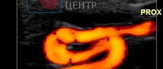
- Ultrasound ultrasound with color mapping (color Doppler mapping)
This ultrasound method is one of the most modern. It not only evaluates blood flow in real time, but also indicates the speed of blood circulation in different color shades. The blood flow directed towards the sensor is colored red, and from the sensor - blue. The brighter the shade, the higher the speed.
- Duplex sonography
Combines duplex scanning and color mapping. As a result, 2 images appear on the screen: monochrome, which shows the structure of the blood vessels, and color, reproducing the blood flow.
Advantages of the ultrasound machine used
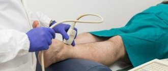
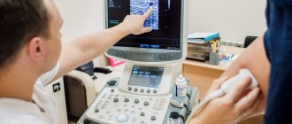
- Ultrasound examination using the Philips HD15 device allows for high-quality and timely diagnosis of diseases at the earliest stages, which facilitates the prescription of competent treatment to achieve a positive result.
- The equipment is compact and portable, while providing high image quality.
- The highly sensitive sensor requires virtually no subsequent adjustments to suit the examination type thanks to automated optimization.
- The device is universal and widely used in practical work.
Indications for the study
It is necessary to do an ultrasound of the veins:
- with varicose veins;
- thrombosis;
- trophic ulcers on the legs;
- the appearance of spider veins;
- thrombophlebitis;
- swelling of the legs;
- varicose veins;
- changes in skin color (usually pink-violet or brown areas appear);
- reducing the volume of one leg;
- frequent seizures;
- frequent goosebumps on the skin;
- tingling, itching, pain, numbness in the legs.
It is also periodically recommended to do vascular ultrasound if there are risk factors:
- with a sedentary lifestyle;
- long standing or sedentary work;
- increased physical activity;
- passion for extreme sports;
- frequent flights;
- hypertension or hypotension;
- overweight;
- constantly wearing high heels and tight underwear;
- presence of bad habits;
- pregnancy.