Magnetic resonance imaging is carried out as an expert method of examination in identifying diseases of the pancreas.
Modern units are able to level out artifacts associated with natural movements (blood flow through vessels, peristalsis, respiratory excursions). The use of phased arrays to enhance the magnetic field in the area of interest allows one to obtain layer-by-layer images of the organs of the biliary system with high contrast resolution. Evaluation of the pancreas is optimized with MR pancreatography demonstrating the ductal system and MR angiography demonstrating the vessels.
The doctor can examine enlarged images of the pathological lesion. Using computer modeling, three-dimensional image reconstruction is available.
Magnetic resonance scanning does not involve invasive penetration into the body, as with endoscopic retrograde cholangiopancreatography (ERCP), and can be performed on an outpatient basis. In the absence of contraindications, the diagnostic procedure is absolutely safe for humans.
Administration of a drug based on gadolinium chelates increases the differences in signal intensity between normal pancreatic parenchyma and altered tissue in malignant tumors. Enhanced MRI is useful in assessing acute pancreatitis and shows the area of necrosis in complicated forms. The study allows you to differentiate hypervascular formations of the pancreas, which can mimic cystic lesions on non-contrast scanning.
Pancreatic diseases
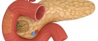
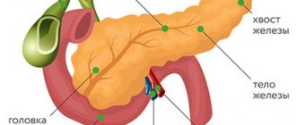
The pancreas is an organ of the digestive and endocrine system. The main functions are the production of pancreatic juice, which contains enzymes, and the production of several important hormones, including insulin. Located posterior to the stomach, closely connected with the duodenum. The pancreas consists of a head, body and tail; blood supply occurs through the pancreaticoduodenal arteries and veins. Most often diagnosed:
- Pancreatitis.
Magnetic resonance imaging for acute organ pathology
Acute inflammation of the pancreas is accompanied by severe pain in the upper abdomen. Other symptoms include nausea, vomiting, diarrhea, flatulence and fever. The cause of acute pancreatitis is gallstones, chronic alcohol consumption, hereditary diseases, injuries, medications, infections, electrolyte and hormonal disorders, high lipid levels, etc. If it is impossible to detect an internal factor, they speak of idiopathic (i.e., self-occurring) form. In the absence of adequate diagnosis and treatment, tissue necrosis occurs.
In acute pancreatitis, MRI shows the presence and degree of necrotization of the pancreas and the area of edema. Magnetic resonance imaging has advantages over CT in diagnosing subtle (initial) changes.
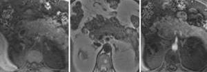
A standard MR scanning protocol of the pancreas, including T2WI, T1WI fat-suppressed and a series of T1WI, GRE sequences before and after gadolinium administration, is a reliable way to determine the stage of acute pancreatitis and the prognosis of the disease.
In severe acute pancreatitis, MRI of the pancreas with contrast is performed to assess parenchymal perfusion and the presence of necrosis. Organ enlargement is clearly visualized on any sequence, and parenchymal edema is better demonstrated on native T1 imaging.
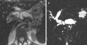
MRI of the pancreas in chronic pancreatitis shows uneven expansion of the main duct with the formation of lateral branches, filling defects
Chronic pancreatitis is a progressive disease associated with the gradual destruction of the pancreas against the background of fibrosis and cellular infiltration, with the development of duct pathology and loss of exocrine and endocrine functions. Clinical manifestations are similar to the acute form. The advanced stage is characterized by underweight, anemia, and the development of type 2 diabetes. With a long-term ongoing process, MRI shows atrophy of the pancreas and pseudocysts. Changes are differentiated from tumor pathology.
- Pancreatic tumors.
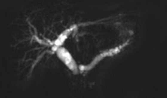
Double duct sign on MRI indicates pancreatic cancer of the head of the organ
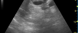
The majority of cases (85%) of malignant lesions are adenocarcinoma of ductal origin with typical localization in the head of the organ. Pancreatic cancer is difficult to detect at an early stage because there are no clinical manifestations. As the pathology progresses, abdominal pain, back pain, weakness, yellowing of the skin and sclera appear. Ultrasonography for tumors smaller than 3 cm shows ambiguous data. Multiparametric MRI with diffusion-weighted imaging and magnetic resonance cholangiopancreatography allow the analysis of morphological changes in the pancreatic parenchyma and duct.
- Cystic and pseudocystic neoplasms.
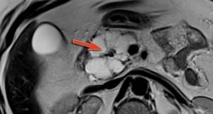
Multicystic lesion of the pancreas on MRI
Cysts and pseudocysts are detected during ultrasound scanning of the abdominal organs; often, when they are small in size, symptoms are nonspecific or absent. It is important to undergo an in-depth study to determine the malignant potential of the tumor. To make a diagnosis, gender, age, and medical history (trauma, alcohol intoxication, chronic diseases of the biliary system, etc.) are important. Final verification is possible after morphological examination.
Signs of cystic neoplasms of the pancreas:
- irregular contour, thickening of the walls;
- presence of partitions;
- visualization of solid components;
- dilated pancreatic duct more than 3 mm;
- calcifications, etc.
Who should not undergo an MRI?
Despite the safety of examination using a magnetic resonance imaging scanner, there are restrictions or complete prohibitions on the procedure. A complete ban on undergoing MRI includes:
- the presence of implants containing metal compounds (cochlear implant, pacemaker, mechanical heart valve);
- the presence in the body of foreign objects with a metal base or inclusions (bullets, fragments);
- presence of tattoos using paint with metals;
- pregnancy period (1st trimester);
- the patient’s condition when artificial life support is required (resuscitation, coma);
- large body weight (more than 120 kg).

The reason for the prohibition of undergoing examination if a patient has permanent metal implants and tattoos on the body is due to the fact that these objects under the influence of the magnetic field of the tomograph can heat up, expand and even move slightly. This situation can bring not only pain, but also injury. Electronic items and metal implants may fail.
It is not a contraindication for magnetic resonance imaging if the patient has dentures or modern braces, as well as titanium implants.
Pregnant patients are prohibited from undergoing the procedure in the 1st trimester, because there is no collected information about the harm or safety of the examination for the correct formation of the fetus.
In some cases, undergoing a magnetic resonance examination is extremely important for the patient's life. According to the doctor's decision, if categorical reasons for refusing the procedure are not found, magnetic resonance imaging can be performed.
It is not advisable to undergo a tomography examination if the following circumstances exist:
- period of breastfeeding and pregnancy (if a contrast agent is used);
- allergy to gadolinium metals included in the contrast agent (extremely rare);
- diagnosed renal or liver failure;
- inability to keep the body still (in some cases, the doctor may suggest medication to induce sleep);
- the presence of mental disorders, acute fears and panic attacks associated with being in the “tunnel” of a tomograph (in this case, you can try to undergo research using open machines);
- severe pain that does not allow you to lie still on the medical table at the time of scanning (the doctor may suggest taking painkillers).
Is MRI performed for diseases of the pancreas?

The reason to sign up for a magnetic resonance examination of the abdominal cavity and retroperitoneal space is the detection in laboratory parameters of changes characteristic of damage to the organs of the biliary system
MRI for diseases of the pancreas is done to identify pathology, determine the stage of the disease and detect complications. The images provide the doctor with valuable information for making a diagnosis. The doctor analyzes:
- location, size and internal structure of the gland;
- zonation of the organ - features of visualization of the body, head and tail;
- involvement of parapancreatic tissue in the pathological process, spread of the tumor to nearby tissues, connection of the neoplasm with the pancreas, damage to the lymph nodes;
- configuration, tumor density, blood supply characteristics, presence of areas of necrosis, calcification (allows to differentiate benign and malignant processes);
- consistency of anastomoses after liver transplantation;
- changes in the parenchyma, the severity of inflammation;
- parasitic lesions;
- developmental anomalies;
- modification of the ducts of the pancreatic and biliary tract as a whole, the presence of strictures, localization, stone formation;
- changes in the retroperitoneal space etc.
Taking into account the absence of long-term consequences after the diagnostic procedure, the study is suitable for dynamic monitoring of cystic neoplasms and monitoring the effectiveness of therapy.
The essence of the method
The principle of contrast is to enhance the displayed object. Haldonium-based preparations are used as a contrast. Galdonium spreads through the bloodstream and accumulates in the tissues of the organ being studied. Contrast enhancement allows you to highlight the clear boundaries of healthy tissue or, conversely, the boundaries of a pathological area. The specialist thus visualizes not only the boundaries, but also the size, structure, and localization of the tumor. It is worth noting that the contrast is completely eliminated from the body and rarely causes an allergic reaction.
MRI of the pancreas: indications

What is better, to do a magnetic resonance or computed tomography, the doctor decides, taking into account the clinical situation and restrictions on the use of the method
There are many indications for MRI of the pancreas. It is recommended to sign up for the procedure if:
- pain in the epigastric region, abdomen, indigestion, inexplicable by tests and ultrasound, appeared, indigestion - foul-smelling stool, unusual color of stool, nausea, vomiting, flatulence, jaundice, etc.;
- family history is burdened - several first-degree relatives have been diagnosed with pancreatic cancer;
- preoperative diagnostics are necessary to study the anatomical features and plan the intervention;
- ultrasound results are ambiguous/suspicious for pancreatic pathology, which implies advanced diagnostics;
- differentiation between cancer and chronic pancreatitis is required;
- deviations from the norm in laboratory parameters indicate disease of the pancreas, gallbladder, ducts, liver;
- there is a suspicion of a tumor in the biliary system, it is necessary to determine the stage of the oncological process, relapse after treatment;
- there are contraindications to computed tomography, including enhanced CT (pregnancy, childhood, allergy to iodine-containing contrast, renal failure, hyperfunction of the thyroid gland);
- There is no effect of therapy for pancreatitis / frequent exacerbations are observed when following a diet (MRI of the pancreas shows choledocholithiasis, intraluminal stones, stage of inflammation);
- it is necessary to exclude postoperative complications, etc.
How is an MRI of the pancreas done?
The greatest diagnostic value is for images obtained on high-field units with a closed contour
Before scanning, you must leave items containing metal in the storage compartment. A cell phone or payment card forgotten in a pocket when exposed to a magnetic field causes distortion in the photo.
After completing the documentation, the x-ray technician escorts the patient to the diagnostic room and places him on the scanner table. The limbs are fixed with soft belts and bolsters, and an intensifying coil is placed above the abdomen.
If an MRI of the pancreas with contrast is planned, a catheter is inserted into the vein. During certain phases of the study, a chelated gadolinium enhancer will be automatically delivered through the drain (using an injector) to improve visualization.
Medical staff monitors the progress of the diagnosis through glass from an adjacent room. Communication takes place via speakerphone; in case of an unforeseen event (an attack of claustrophobia, deterioration in health), the patient has a special signal button at hand. Technical noise produced by equipment can be leveled out using headphones.
After the native series of images, there is a wait for the dye to spread through the bloodstream to the organs of the biliary tract. The duration of MRI with contrast is 30-45 minutes. The results will be ready within an hour. There are no obstacles to daily activities.
Progress of the procedure
The patient takes off his watch, jewelry, and puts on disposable clothing (you can leave your clothes if they do not have metal parts). During the procedure you should not have your phone or credit cards with you.
During the examination, the patient is in a supine position on the tomograph table. The table slides into the tunnel (if the device is closed). It is mandatory to remain still during the procedure, which lasts from 30 minutes. up to an hour.
To create comfort, there is a constant supply of fresh air into the tunnel, lighting, and the ability to use earplugs to eliminate the loud sound of the tomograph. The doctor has the opportunity to observe the patient during the procedure and communicate with him via an intercom.
When planning to use a contrast agent, before starting the study, a subcutaneous test is performed to determine the patient's sensitivity to it. In the absence of allergic manifestations, contrast is administered intravenously during the study.
MRI of the pancreas, how to prepare?

Ask your doctor if you have any questions about MRI scans.
Magnetic resonance imaging is often performed routinely; in urgent situations with an acute abdomen, it is preferable to do a CT scan. Preparation for an MRI of the pancreas implies:
- Obtaining a referral for research. The document must indicate the intended diagnosis, area/s of interest, type - with or without contrast;
- Dieting. A few days before the procedure, products containing starch, yeast, sugar - baked goods, confectionery - are excluded from the diet. Excessive flatulence and fermentation processes in the intestines are caused by fresh vegetables (cabbage, peas, beans), fruits, herbs, mushrooms, milk, brown bread, etc. Alcohol, kvass and drinks with gas can cause the movement of small stones and accompanying inflammation, which will provoke swelling of the gland and gallstone colic. You should avoid fatty meats, fish, smoked meats, marinades, and spices. A 6-hour fast is required before the test. The paramagnetic gadolinium and prolonged lying on the back can cause autonomic reactions in the form of dizziness, nausea, and drooling. You can prevent such phenomena by having a light snack 30 minutes before leaving the house.
- Taking medications. Medicines for routine therapy are taken as usual. Antispasmodics, enzymes, adsorbents, laxatives are used exclusively on the recommendation of a doctor. An enema is needed for chronic constipation (if it is planned to examine all organs of the gastrointestinal tract).
Don’t forget to take with you the results of ultrasound, FGDS, images and descriptions of previously performed MRI/CT, epicrisis from the hospital, conclusion of the oncoconsilium. If the study is paid for by an insurance company, a compulsory medical insurance or voluntary health insurance policy will be required.
How is tomography performed?
Before starting the examination, the patient must take a lying position inside the tomograph. Then the subject is injected with a contrast agent if the scanning is carried out using a contrast protocol. In some cases, during the contrast procedure, you may experience a feeling of nausea, vomiting, malaise or weakness. You should immediately report these symptoms to your doctor, who will promptly take measures to stop the allergic reaction to the contrast agent.
The scanning procedure itself takes an average of 3-7 minutes, during which the subject must lie as still as possible and follow the operator’s commands to hold his breath. The radiologist will need another 30-40 minutes to decipher the diagnostic data. The result of the CT examination will be a written conclusion from the doctor and recording of the tomograms on electronic or film media.
What can you eat before an MRI of the pancreas?

It is preferable to prepare dishes using gentle heat treatment - boiling, baking, stewing
To ensure that food is fully digested, the following are allowed on the eve of the procedure:
- lean meat, dietary fish, skinned chicken breast;
- baked vegetables and fruits;
- porridge with water;
- omelettes;
- unrich broths;
- crackers, dry cookies;
- vegetable oil in small quantities;
- unsweetened compotes, still water, herbal teas, diluted juices without pulp.
Preparation
For a high-quality diagnosis, the examinee must provide the doctor with all available medical information related to the pathology:
- excerpts from case histories;
- ultrasound, x-ray, tests for tumor markers;
- direction for scanning.
If a multislice CT scan is performed with contrast, patients with kidney disease will need to have a blood test for creatinine. It will help specialists decide whether to perform a CT scan with contrast.
An allergy test is a very important aspect of preparation for contrast MSCT of the pancreas. It is carried out to avoid anaphylactic reactions to iodine-containing contrast material directly in the medical clinic before the examination.
You need to prepare for a CT scan of any organs of the abdominal cavity and retroperitoneal space. Three to two days before the tomography, you should start following a diet that helps reduce gas formation. The patient should give up fresh vegetables and fruits, cabbage, legumes, baked goods, baked goods and other high-fiber foods. The evening before the study, no later than 21:00, a light dinner is allowed, after which food intake should be stopped. After dinner, you can take two tablets of espumisan or activated carbon to reduce gas formation. Tomography of the pancreas is performed on an empty stomach. The minimum fasting period should be at least 6 hours. On the day of the study, 30-40 minutes before the start of MSCT, you need to take two No-Spa tablets.
The issue of preparing for a CT scan of the pancreas should be approached responsibly. If diet requirements are not followed, CT scan results may be incorrect.
Normal size of the pancreas on MRI
Organ parameters are variable:
| Department | Dimensions (mm) |
| Head | 11- 35 |
| Body | 4- 30 |
| Tail | 7- 30 |
The concept of normal on magnetic resonance images is relative. Sizes depend on gender, height, age, food load. A slight deviation from generally accepted indicators, provided the structure of the organ parenchyma is normal and functions are intact, is not considered a pathology. The length of the pancreas is 15-23 cm, width 4.5-6 cm, weight about 70-80 grams.
Normally, the organ is not enlarged, the contours are clear, even, and the dimensions correspond to the person’s build. The pancreatic duct is not dilated, the surrounding tissue is without any features. No changes in the MR signal were detected.