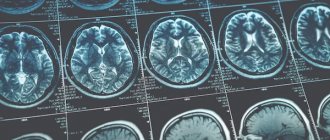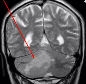
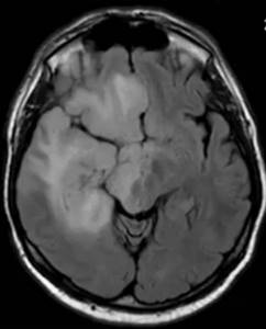
Cerebellar tumor on MRI (indicated by arrow) Brain tumors have long been diagnosed solely on clinical grounds, often too late. With the advent of neuroimaging methods, it has become possible to identify dangerous pathologies in the early stages of their development. Already in the first stages of formation, a brain tumor is clearly visible on MRI, and doctors can plan further actions. Early diagnosis increases the chances of recovery even with cancer.
Symptoms of brain cancer
Malignant tumors account for almost half of the identified intracranial neoplasms. Common manifestations of pathological changes in the brain can be:
- headaches, often in the morning, with nausea and vomiting;
- vertigo (dizziness of central origin);
- impaired coordination of movements;
- deterioration of vision, hearing;
- speech disorders;
- irritability;
- drowsiness;
- convulsions;
- behavioral disorders;
- weakness in the muscles of the face or limbs;
- numbness, paresthesia in certain parts of the body;
- mental disorders, etc.
The clinical picture of tumors differs depending on the location, size, and type of tumor. Pathology is differentiated based on data obtained from instrumental and laboratory examinations and histological analysis of tissues.
Based on how a brain tumor looks on an MRI, doctors make assumptions about its nature. There can be no unambiguous statements, since the verification of formations is carried out using histological analysis. Characteristic signs of malignant brain tumors on MRI images are:
- their shape is incorrect;
- unclear contours of neoplasms;
- pronounced perifocal edema;
- mass effect (displacement of nearby structures);
- hydrocephalus;
- heterogeneous accumulation of contrast (sometimes they do not accumulate at all);
- “variegation” of the tumor (hemorrhages, necrosis and/or cysts in the structure), etc.
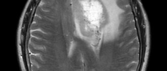
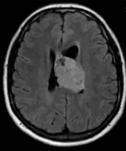
Glioblastoma on brain MRI without contrast
Benign and malignant tumors
Before moving on to the appearance of tumors, let’s figure out what the fundamental difference between these two formations is:
- Benign tumor: Slow growing
- Incapable of creating metastases, that is, spreading throughout the body
- Easy to treat
- May develop into malignant with further enlargement and if located close to internal organs
- Grows fast
However, it is worth remembering that the division is quite arbitrary, and these characteristics are not always strictly observed, but most often this is the case. To determine the nature of the tumor, its size, and stage of development, it is optimal to perform an MRI with contrast. What visual markers do these tumors have on MRI?
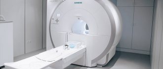
MRI in our center
Power 1.5 Tesla
High image quality
Study for patients up to 250 kg
Burn to disc for free
Causes of brain tumors
The exact mechanism of formation of malignant tumors has not been established. Risk factors include age over 70 years and being male. More often, tumor pathologies of the brain are found in patients who have:
- family history;
- contacts with carcinogenic substances;
- radiation exposure;
- history of traumatic brain injury;
- past inflammatory pathologies of the brain.
In some patients with detected tumors, doctors are unable to identify a single provoking factor, while in people who are regularly exposed to them, there are no brain tumors. Such information casts doubt on existing hypotheses about the origin of oncopathologies of the central nervous system (CNS).
Ependymoma on MRI scan of the brain
Head tumors are often secondary in nature, i.e. metastases. According to localization, formations can be located in the bones of the skull, soft and dura maters, in gray and white matter. Melanomas, cancers of the kidney, prostate, lung, breast, and stomach most often metastasize to the structures of the head.
Brain metastases on post-contrast images (indicated by arrows)
Tests and examinations for cancer: myths and reality
Content:

Can cancer be detected by a blood or urine test? Is it possible to diagnose cancer using a saliva test? Do I need to undergo CT and MRI scans regularly? Is it worth getting tested for tumor markers? Together with Olga Aleksandrovna Goncharova, an oncologist of the highest category at the Consultative and Diagnostic Center of the National Medical Research Center of Oncology named after. N.N. Petrov, we figured out the effectiveness and feasibility of popular methods for early diagnosis of cancer.
– Olga Alexandrovna, is it possible to detect cancer using a blood test?
– It is impossible to determine cancer from a blood test. Of course, certain blood test indicators can alert the attending physician and the patient. For example, low hemoglobin, high ESR, increased bilirubin, high ALT, AST, etc. Such analysis results should serve as a reason for further in-depth examination.
– What about urine analysis?
– It’s also impossible, but some changes in the urine test should alert you. For example, the appearance of red blood cells can indicate both a tumor of the kidneys, bladder, ureter, and diseases of the genitourinary system unrelated to cancer pathology. That is, these changes in clinical urine analysis should serve as a reason for further examination of the genitourinary system.

– Is it possible to diagnose cancer using a saliva test?
– Unfortunately, diagnosing cancer using saliva examination is currently impossible. At the University of California, a team of specialists led by David Wong, MD, suggested that traces of RNA can be found in human saliva, which can be used to determine the presence of tumor cells in the body. In 2015, they published the results of their study, which partially confirmed their hypothesis. However, these tests are still under development, and it cannot be said with certainty that saliva can be used to diagnose cancer.
– Can tumor marker tests detect cancer?
- No. Tumor markers are used to monitor existing malignant tumors. They are criteria for the so-called tumor response to treatment, when relapse or metastasis of an already treated tumor is detected. This is not at all a tool for screening and early diagnosis of cancer. Tumor markers are substances, often of a protein nature, that are released either by the tumor itself or by the body in response to its development. Due to the specificity of each protein, it is possible to predict the focus of the disease, the damage to certain organs at different tumor locations. But we must remember that an increase in the level of tumor markers is not a 100% sign of cancer. They can increase even in the absence of cancer for many other reasons: due to inflammatory processes, infections, pregnancy, benign tumors and taking certain medications. You also need to understand that normal levels of tumor markers cannot guarantee the absence of malignant neoplasms. And not all cancer patients diagnosed with malignant neoplasms have elevated levels of tumor markers. What is important is not a single tumor marker result, but monitoring. Its concentration level makes it possible to determine the onset of changes in the formation of tumor recurrence or metastasis 1-6 months faster than the use of other diagnostic methods.
– Is it possible to detect cancer at an early stage using regular MRI and PET/CT scans?
– Unfortunately, not either. MRI and PET/CT are not used anywhere in the world for cancer screening. There are several reasons. Firstly, during such a study it is often possible to obtain a false positive or false negative result, which we often encounter in our practice. Secondly, undergoing only one type of research is not informative. Diagnosing the disease requires an integrated approach. And thirdly, the annual completion of such examinations carries additional radiation exposure. MRI and PET/CT should only be performed as prescribed by a doctor. It is the specialist who can clearly determine the purpose of the study, indications and contraindications.
– Why can CT, MRI and other research methods produce incorrect results?
– Sometimes false positive results can be associated with inflammatory processes or other non-oncological pathologies in the body. Sometimes there is a misinterpretation of these studies. Excited patients often come to us with the results of a CT or MRI scan they have already performed. We review discs at our institution, and the conclusions of the studies provided do not always coincide with the opinions of our experts. But it happens the other way around: the pictures show early oncological pathology, but this is not indicated in the medical report. Correct assessment of the results of MRI, CT, X-ray or mammography is the key to a correct diagnosis. If you suspect an oncological pathology, it is recommended to undergo examinations at a specialized oncological institution. This significantly reduces the risk of misinterpretation of CT and MRI results, and also avoids unnecessary and uninformative studies.
It is worth noting that early diagnosis of cancer is possible only with a combination of various research methods. A specialist can determine what kind of research is needed for each specific person. If you are concerned about the risk of developing cancer, you should first consult an oncologist. In the next article we will tell you which methods of early cancer diagnosis are really effective.
Bibliography:
1. “Organization of preventive medical examination and medical examination of certain groups of the adult population. Methodological recommendations" (approved by the Ministry of Health of Russia on October 22, 2019) 2. Clinical recommendations "Breast cancer". Approved at a meeting of the scientific and practical council of the Ministry of Health of the Russian Federation, 2021 3. Clinical recommendations “Malignant neoplasms of the colon and rectosigmoid region.” Approved at a meeting of the scientific and practical council of the Ministry of Health of the Russian Federation, 2021 4. Clinical recommendations “Malignant neoplasms of the bronchi and lung.” Approved at a meeting of the scientific and practical council of the Ministry of Health of the Russian Federation, 2021 5. Radiation diagnostics of the chest organs. National leadership. Under. Ed. S.K.Ternova, Moscow, 2014. 6. Polidanov M.A., Blokhin I.S. Computed and magnetic resonance imaging: basic principles, procedure and distinctive features. Petrozavodsk, 2020.
Can an MRI show a brain tumor?
Based on the results of magnetic resonance imaging, doctors can identify changes in tissue structure. Brain tumors on MRI are determined by direct and indirect signs. Doctors suspect a pathological formation based on the type and uniformity of changes in the MR signal:
- hypo- (weaker than surrounding areas) or hyperintense (response stronger than from normal areas);
- heteromodified (different characteristics are mixed);
- isointense (the tumor looks like other tissues, but differs from the normal architecture of the brain).
There are also indirect signs. The latter give reason to suspect a brain tumor against the background of an isointense tissue response or confirm its presence with an altered MR signal.
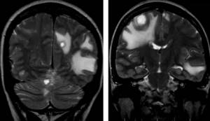
Multiple metastases in the brain substance on MRI without contrast
Secondary manifestations of pathology may be:
- lateral dislocation of the median structures;
- displacement of the vascular bundle;
- changes in the size and deformation of the ventricles;
- axial dislocation;
- occlusive hydrocephalus due to blockage of the cerebrospinal fluid pathways;
- displacement or deformation of the basal cisterns;
- cerebral edema in the tumor area and around it (local, generalized, total, periventricular).
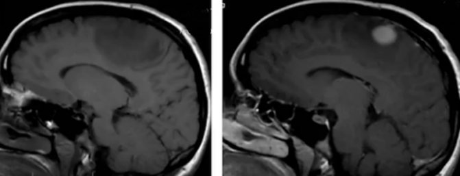
Pre- and post-contrast images of a malignant brain tumor
Using MRI for brain cancer, the localization of the tumor, the degree of its invasion into neighboring structures, features of the blood supply, stage of the disease and the presence of metastases are revealed. To obtain additional information, scanning is carried out in various sequences (T1, T2, FLAIR, diffusion-weighted, etc.), in native mode and after contrast.
How safe is the procedure?
Can I do an MRI for oncology? - a question that interests all patients who are undergoing such a procedure.
The answer is simple: the safety of MRI, including when performed multiple times, regardless of the stage of development of the oncological process, has been proven. The method can also be used if there is a suspicion of the presence of a neoplasm in the body, since tomography does not accelerate the process of tumor cell division and does not cause the degeneration of a benign process into a malignant one.
Undergoing examination through exposure to electromagnetic pulses does not harm the body, since tomography does not involve the presence of ionizing radiation or X-rays, as is the case with CT.
Note!
People often confuse MRI and CT (there is an opinion that computed tomography, by affecting a weakened body, only accelerates the growth of tumors), which causes unreasonable fears.

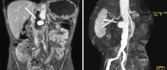
What does MRI of the abdominal cavity and retroperitoneal space show?
The abdominal cavity contains the organs of the digestive system and the endocrine glands. MRI helps to examine the structure of parenchymal organs: the liver with ducts, pancreas, spleen, retroperitoneal kidneys and adrenal glands, as well as lymph nodes and visceral fat. Hollow organs (esophagus, stomach, intestinal loops) are not assessed by MRI.
MRI is prescribed in the following situations:
- stones in the biliary system - MRI will show the presence of stones in the cavity of the gallbladder, common bile duct or bile ducts. A special mode - MR cholangiography is used in diagnosing the causes of obstructive jaundice;
- detection of primary hepatocellular cancer, as well as determination of the spread of metastases from oncological processes of another localization;
- inflammatory liver diseases of various nature (viral, toxic, autoimmune hepatitis), the presence of fibrotic or cirrhotic restructuring;
- diagnosis of pancreatic diseases of inflammatory or tumor origin with identification of cystic degeneration, areas of destruction and assessment of the patency of the common pancreatic duct;
- study of the structure of the spleen, the presence of cysts, various tumors and traumatic injuries;
- inflammatory kidney diseases (pyelonephritis, glomerulonephritis, kidney carbuncle);
- violation of the outflow of urine from the kidneys - MRI shows the location of the kidney block and helps determine its nature (calculus, tumor).
To increase the diagnostic information, an MRI of the abdominal cavity is performed on an empty stomach or 4 hours after eating or drinking.
What does an MRI of the spine show?
MRI allows you to study the structure of individual vertebrae, intervertebral discs, the spinal cord, spinal nerve roots, as well as joints and ligaments of the spinal column. Diagnostics is divided into zones - cervical, thoracic, lumbar and sacrococcygeal sections; you can examine one section or several at once. Tomography allows you to detect:
- signs of a degenerative-dystrophic process in the spine (osteochondrosis, spondylosis, spondyloarthrosis);
- protrusion of intervertebral discs and herniation;
- narrowing of the lumen of the spinal canal, as well as compression of the roots of the spinal nerves by hernias or neoplasms;
- improper development of the spinal cord and bone tissue of the spinal column;
- activity of demyelinating diseases, incl. multiple sclerosis;
- inflammatory processes and tumors.
What do spinal metastases look like on MRI?
The results of MR scanning are layer-by-layer images of the area under study in several projections. The radiologist has the opportunity to examine any area of the spine and identify the slightest deviations from the norm. Pathological processes are manifested by changes in impulses from tissues. The signal can be hypo-, iso- and hyperintense.
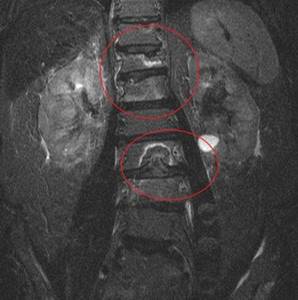
Secondary focal lesion of the spine (zones of destruction are highlighted)
Depending on the pulse sequence protocol used, the radiologist makes conclusions about the possible origin of the unusual MR signal.
More often, tumors of the breast in women, prostate tumors in men, and lungs metastasize to the spine. Somewhat less frequently, lymphoma, melanoma and renal cell carcinoma are screened into this area. The bodies of the lower thoracic and lumbar vertebrae are most often affected.
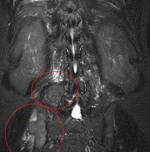
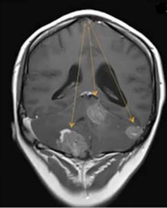
MRI scan: at the top there is a large paravertebral formation, at the bottom there is a secondary lesion of the pelvic bones
Secondary tumors of various types are differently visible on MRI images:
- osteolytic metastases. Contrast agent accumulates well. T1-weighted images show a hypo- or isointense signal. That is, they look darker than the surrounding tissues or match them. When using T2 sequences, they give an iso- or hyperintense signal. Osteolytic metastases are clearly visible in the mode with suppression of the signal from adipose tissue. These formations on the images look like hyperintense lesions;
- osteoblastic metastases. Poorly accumulates contrast agent. T1 and T2 weighted images appear dark. When the signal from adipose tissue is suppressed, they appear as iso- or slightly hyperintense areas;
- secondary tumors of mixed nature. T1 sequences can produce both hypo- and hyperintense signals.
The sensitivity of the method is about 90%. However, an MRI scan only confirms or suggests a diagnosis. Based on tomography results, it is impossible to distinguish metastases from primary spinal tumors. For the purpose of differential diagnosis, aspiration or conventional biopsy is used, followed by cytological and histological examination.
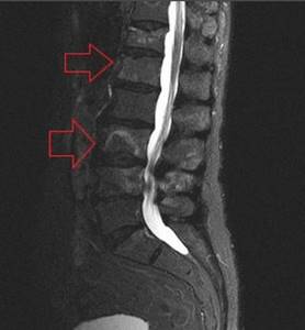
Metastases in the spine on MRI (indicated by arrows)
Features of MRI
Important features of MRI include:
- High accuracy of examination results. An MRI can provide a three-dimensional image of the internal organs of various parts of the body. Thanks to this, the doctor can quickly detect the emergence and development of pathology at the earliest stages, when there are still practically no clinical manifestations of the disease;
- Lack of complex preparation. Carrying out an MRI does not require any special preparation. In the event that an examination of the abdominal cavity and pelvic organs is carried out, you will have to temporarily exclude from the diet foods that lead to increased gas formation. And when diagnosing the pelvic organs, you need to fill the bladder in advance;
- Affordable price. The cost of an MRI examination is at a level that is quite affordable for many residents of our country. Of course, prices in Russian clinics can vary greatly, since different equipment is used to diagnose the condition of soft tissues, cartilage and brain structures.
What can an MRI show?
The unique capabilities of tomography are due to the phenomenon of magnetic resonance of hydrogen protons used in it. Under the influence of a high-voltage magnetic field, the nuclei are excited, and the electromagnetic responses received from them are recorded by the apparatus. After special software processing, images of the internal structure of organs are obtained. However, the structural features of organs and tissues are not all that the MRI method shows. Thanks to the magnetic field and electromagnetic resonance from protons, this technique allows you to estimate the number of hydrogen atoms, and therefore liquid, in various organs and tissues, i.e. visualizes their chemical composition. The diagnostic study is carried out on a 1.5 Tesla tomograph Siemens Symphony installed in 2015, which allows for expert-class diagnostics.
What does an MRI of the soft tissues of the neck show?
MRI of organs and soft tissues located in the neck area makes it possible to study the structure of the thyroid and salivary glands, larynx, lymph nodes, muscles and fascial spaces located prevertebrally, i.e. in front of the spinal column. Doctors recommend MRI to diagnose the following diseases:
- thyroid neoplasms - tomography shows the boundaries, size of the tumor, and also helps to identify screenings of the oncological process beyond the thyroid tissue - into the cervical lymph nodes or neighboring organs;
- inflammation and tumors of the salivary glands, the presence of stones in the ducts;
- lymphadenopathy of the neck - enlarged lymph nodes in the neck can be associated with a chronic or acute inflammatory process in the oral cavity, ENT organs, and also appears with cancer and lymphoproliferative diseases. MRI helps in differential diagnosis and establishing the cause of lymph node damage;
- neoplastic processes of the pharynx and larynx - tomography will help doctors decide on treatment tactics, and is also used to monitor the effectiveness of the therapy.